
L8793
Anti-MAP1LC3A antibody
rabbit polyclonal
Synonyma:
Anti-MAP1 light chain 3-like protein 1, Anti-MAP1A/1B light chain 3 A, Anti-MAP1A/MAP1B LC3 A, Anti-MAP1ALC3, Anti-MAP1BLC3, Anti-MAP1LC3A, Anti-Microtubule-associated protein 1 light chain 3 alpha
O této položce
Název produktu
Anti-LC3A antibody produced in rabbit, ~1 mg/mL, affinity isolated antibody, buffered aqueous solution
biological source
rabbit
Quality Level
conjugate
unconjugated
antibody form
affinity isolated antibody
antibody product type
primary antibodies
clone
polyclonal
form
buffered aqueous solution
mol wt
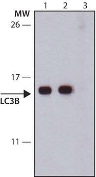
antigen 16-18 kDa
species reactivity
mouse, human, rat
concentration
~1 mg/mL
technique(s)
immunoprecipitation (IP): 5-10 μg using extracts of human U87 cells, western blot: 0.5-1 μg/mL using whole extracts of rat and mouse brain
UniProt accession no.
shipped in
dry ice
storage temp.
−20°C
target post-translational modification
unmodified
Gene Information
human ... MAP1LC3A(84557)
mouse ... Map1lc3a(66734)
rat ... Map1lc3a(362245)
General description
Immunogen
Application
- immunohistochemistry
- immunostaining
- western blotting[1]
Biochem/physiol Actions
Physical form
Preparation Note
Disclaimer
1 of 1
Tato položka | |||
|---|---|---|---|
| conjugate unconjugated | conjugate unconjugated | conjugate unconjugated | conjugate - |
| Quality Level 200 | Quality Level 200 | Quality Level 200 | Quality Level - |
| antibody form affinity isolated antibody | antibody form affinity isolated antibody | antibody form purified immunoglobulin | antibody form purified immunoglobulin |
| biological source rabbit | biological source rabbit | biological source mouse | biological source rabbit |
| species reactivity mouse, human, rat | species reactivity mouse, human, rat | species reactivity human | species reactivity human, mouse |
| UniProt accession no. | UniProt accession no. | UniProt accession no. | UniProt accession no. |


Still not finding the right product?
Explore all of our products under Anti-LC3A antibody produced in rabbit
— nebo —
Vyzkoušejte náš nástroj Nástroj pro výběr produktů a zúžte své možnosti.
Skladovací třída
10 - Combustible liquids
wgk
WGK 3
flash_point_f
Not applicable
flash_point_c
Not applicable
ppe
Eyeshields, Gloves, multi-purpose combination respirator cartridge (US)
Vyberte jednu z posledních verzí:
Již tento produkt vlastníte?
Dokumenty související s produkty, které jste v minulosti zakoupili, byly za účelem usnadnění shromážděny ve vaší Knihovně dokumentů.
Související obsah
Instructions
Globální číslo obchodní položky
| Skladová položka | GTIN |
|---|---|
| L8793-200UL | 04061838031556 |