


Cells are thawed and expanded in RPMI-1640 (Product No. R0883), 10% FBS (Product No. ES-009-B), 1X L-Glutamine (Product No. TMS-002-C), 1X non-essential amino acids (Product No. TMS-001-C), 1X HEPES Buffer Solution (Product No. TMS003-C) and 0.0054X β-Mercaptoethanol (Product No. ES-007-E).
SCC142M
DC2.4 Mouse Dendritic Cell Line
Mouse
Synonim(y):
DC 2.4 Cell Line
Zaloguj się, aby wyświetlić ceny organizacyjne i kontraktowe.
Wybierz wielkość
Zmień widok
| Gabaryty przesyłki | SKU | Dostępność | Cena netto |
|---|---|---|---|
| 1 vial | Skontaktuj się z Obsługą Klienta, aby uzyskać informacje na temat dostępności | 7580,00 zł |
Informacje o tej pozycji
UNSPSC Code:
41106514
NACRES:
NA.81
eCl@ss:
32011203
Biological source:
mouse
7580,00 zł
Skontaktuj się z Obsługą Klienta, aby uzyskać informacje na temat dostępności
Pomoc techniczna
Potrzebujesz pomocy? Nasz zespół doświadczonych naukowców chętnie Ci pomoże.
Pozwól nam pomócNazwa produktu
DC2.4 Mouse Dendritic Cell Line, DC2.4 dendritic cell line can be used to study dendritic cell biology and immune response.
biological source
mouse
Quality Level
technique(s)
cell culture | mammalian: suitable
General description
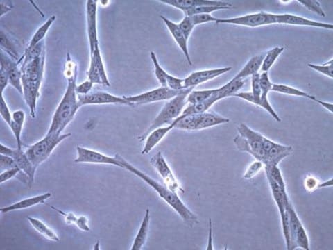
.4 are immortalized murine dendritic cells created by transducing bone marrow isolates of C57BL/6 mice with retrovirus vectors expressing murine granulocyte-macrophage CSF (GM-CSF) and the myc and raf oncogenes [1]. DC2.4 exhibits characteristic features of dendritic cells including cell morphology and the expression of dendritic cell-specific markers and the ability to phagocytose and present exogenous antigens on both MHC class I and class II molecules [1].
Dendritic cells (DC) are the antigen presenting cells of the immune system and are found in most tissues, particularly those that are in contact with the external environment (e.g., skin and the inner linings of the nose, lungs, stomach and intestine). First described in 1973 [2], one of the primary functions of DCs is to phagocytose foreign pathogens and present the processed antigens to naïve T cells to regulate adaptive immune responses. DCs also express Toll-like receptors and help regulate the innate immune responses. Despite their distribution in most tissues, DC are present at low numbers in vivo and are difficult to maintain in vitro. These difficulties have limited the studies of dendritic cells.
Application
Research Category
Immune Response
Inflammation & Immunology
Immune Response
Inflammation & Immunology
Subject to local law, this product is intended to be sold for internal in vitro research use only subject to terms and conditions found here: www.sigmaaldrich.com/restrictedcelluse. This product may not be: re-engineered or copied; used to make derivatives, modifications or functional equivalents; used to obtain patents or other IP claiming use of the product; used to develop, test, or manufacturer a commercial product; used as a component in a commercial product; resold or licensed; used in any clinical applications or trials; or used in humans. A license or limited commercial use agreement is required for use by any for-profit entity, use in services, and use in sponsored academic research. For information regarding any such use, please contact [email protected].
Biochem/physiol Actions
Dendritic Cell Line
Preparation Note
Store in liquid nitrogen. The cells can be cultured for at least 10 passages after initial thawing without significantly affecting the cell marker expression and functionality.
Analysis Note
• Each vial contains ≥ 1X10⁶ viable cells.
• Cells are tested negative for infectious diseases by a Mouse Essential CLEAR panel by Charles River Animal Diagnostic Services.
• Cells are verified to be of mouse origin and negative for inter-species contamination from rat, chinese hamster, Golden Syrian hamster, human and non-human primate (NHP) as assessed by a Contamination Clear panel by Charles River Animal Diagnostic Services.
• Cells are negative for mycoplasma contamination.
Ta strona może zawierać tekst przetłumaczony maszynowo.
1 of 1
Ta pozycja | |||
|---|---|---|---|
| technique(s) cell culture | mammalian: suitable | technique(s) cell culture | mammalian: suitable | technique(s) cell culture | mammalian: suitable | technique(s) cell culture | mammalian: suitable |
| biological source mouse | biological source mouse | biological source mouse | biological source human |
| Quality Level 100 | Quality Level - | Quality Level - | Quality Level 100 |
Klasa składowania
12 - Non Combustible Liquids
wgk
WGK 1
flash_point_f
Not applicable
flash_point_c
Not applicable
Certyfikaty analizy (CoA)
Poszukaj Certyfikaty analizy (CoA), wpisując numer partii/serii produktów. Numery serii i partii można znaleźć na etykiecie produktu po słowach „seria” lub „partia”.
Masz już ten produkt?
Dokumenty związane z niedawno zakupionymi produktami zostały zamieszczone w Bibliotece dokumentów.
Powiązane treści
R M Steinman et al.
The Journal of experimental medicine, 137(5), 1142-1162 (1973-05-01)
A novel cell type has been identified in adherent cell populations prepared from mouse peripheral lymphoid organs (spleen, lymph node, Peyer's patch). Though present in small numbers (0.1-1.6% of the total nucleated cells) the cells have distinct morphological features. The
Z Shen et al.
Journal of immunology (Baltimore, Md. : 1950), 158(6), 2723-2730 (1997-03-15)
Pathways for presenting proteins from the extracellular fluids on MHC class I molecules have been described in macrophages. However, it is uncertain whether similar mechanisms exist in dendritic cells, because conventional preparations of these cells can be contaminated with macrophages.
Vinit Sheth et al.
ACS nano, 17(9), 8376-8392 (2023-04-19)
Super-resolution microscopy can transform our understanding of nanoparticle-cell interactions. Here, we established a super-resolution imaging technology to visualize nanoparticle distributions inside mammalian cells. The cells were exposed to metallic nanoparticles and then embedded within different swellable hydrogels to enable quantitative
Numer pozycji handlu globalnego
| SKU | NUMER GTIN |
|---|---|
| SCC142 | 04054839344213 |
-
Hello, I wanted to know what culture media should be used for this cell line.
1 answer-
Helpful?
-