
MABT366

Anti-Claudin-1/CLDN1 Antibody, clone 7A5
clone 7A5, from mouse
Synonim(y):
Claudin-1, Senescence-associated epithelial membrane protein
Wybierz wielkość
| Gabaryty przesyłki | SKU | Dostępność | Cena netto |
|---|---|---|---|
| 100 μg | Przewidywany termin wysyłki12 maja 2026zKuehne + Nagel Sp. z o.o. | 1910,00 zł |
Informacje o tej pozycji
1910,00 zł
Przewidywany termin wysyłki12 maja 2026Szczegóły
biological source
mouse
Quality Level
conjugate
unconjugated
antibody form
purified immunoglobulin
antibody product type
primary antibodies
clone
7A5, monoclonal
species reactivity
human
should not react with
mouse
technique(s)
flow cytometry: suitable, immunocytochemistry: suitable, western blot: suitable
isotype
IgG1κ
NCBI accession no.
UniProt accession no.
shipped in
dry ice
target post-translational modification
unmodified
Gene Information
human ... CLDN1(9076), NISCH(11188)
General description
Immunogen
Application
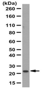
Western Blotting Analysis: 0.5 µg/mL from a representative lot detected exogenously expressed claudin-1 in CLDN1-transfected, but not mock-transfected, HT1080 cells (Courtesy of Professor Masuo Kondoh, PhD, Osaka University, Japan).
Immunocytochemistry Analysis: A representative lot immunostained the surface of Huh-7.5.1 human hepatoma cells, but not the non-claudin-1-/CLDN1-expressing S7-A cells (Fukasawa, M., et al. (2015). J. Virol. 89(9):4866-4879).
Flow Cytometry Analysis: A representative lot specifically immunostained HT1080 cells expressing exogenously transfected human claudin-1 (CLDN1), but not HT1080 cells expressing human claudin-2, -4, -5, -6, -7, or -9, nor L cells expressing mouse claudin-1 (Fukasawa, M., et al. (2015). J. Virol. 89(9):4866-4879).
Flow Cytometry Analysis: A representative lot immunostained HEK293T transfectants expressing FLAG-tagged human claudin-1/CLDN1 and human-mouse claudin-1 chimeras with the second human extracellular loop (EL2), but not chimeras with the mouse EL2. M152L, but not V155I, mutation in human EL2 abolished the immunoreactivity (Fukasawa, M., et al. (2015). J. Virol. 89(9):4866-4879).
Western Blotting Analysis: A representative lot detected FLAG-tagged human claudin-1/CLDN1 and human-mouse claudin-1 chimeras with the second human extracellular loop (EL2), but not chimeras with the mouse EL2. M152L, but not V155I, mutation in the second human extracellular loop abolished the immunoreactivity (Fukasawa, M., et al. (2015). J. Virol. 89(9):4866-4879).
ELISA Analysis: A representative lot detected claudin-1/CLDN1 immunoreactivity in 3.7% formaldehyde-fixed Huh-7.5.1 human hepatoma cells by "cell ELISA" (Fukasawa, M., et al. (2015). J. Virol. 89(9):4866-4879).
Neutralization Analysis: A representative lot inhibited HCV infection of cultured Huh-7.5.1 human hepatoma cells in vitro and of human liver-chimeric mice in vivo (Fukasawa, M., et al. (2015). J. Virol. 89(9):4866-4879).
Cell Structure
Infectious Diseases - Viral
Biochem/physiol Actions
Physical form
Preparation Note
Handling Recommendations: Upon receipt and prior to removing the cap, centrifuge the vial and gently mix the solution. Aliquot into microcentrifuge tubes and store at -20°C. Avoid repeated freeze/thaw cycles, which may damage IgG and affect product performance.
Analysis Note
Immunocytochemistry Analysis: 10 µg/mL of this antibody detected Claudin-1/CLDN1 in HepG2 cells.
Other Notes
Disclaimer
1 of 1
Ta pozycja | |||
|---|---|---|---|
| biological source mouse | biological source rabbit | biological source rabbit | biological source mouse |
| Gene Information human ... CLDN1(9076), NISCH(11188) | Gene Information human ... CLDN7(1366) | Gene Information human ... CLDN11(5010) | Gene Information human ... CLDN2(9075) |
| antibody form purified immunoglobulin | antibody form affinity isolated antibody | antibody form affinity isolated antibody | antibody form purified immunoglobulin |
| conjugate unconjugated | conjugate unconjugated | conjugate unconjugated | conjugate unconjugated |
| clone 7A5, monoclonal | clone polyclonal | clone polyclonal | clone 3F1, monoclonal |
| species reactivity human | species reactivity human, rat, mouse | species reactivity human | species reactivity human |


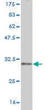
Still not finding the right product?
Wypróbuj nasze narzędzie Narzędzie selektora produktów, aby zawęzić opcje.
Klasa składowania
12 - Non Combustible Liquids
wgk
WGK 2
flash_point_f
Not applicable
flash_point_c
Not applicable
Certyfikaty analizy (CoA)
Poszukaj Certyfikaty analizy (CoA), wpisując numer partii/serii produktów. Numery serii i partii można znaleźć na etykiecie produktu po słowach „seria” lub „partia”.
Masz już ten produkt?
Dokumenty związane z niedawno zakupionymi produktami zostały zamieszczone w Bibliotece dokumentów.
Numer pozycji handlu globalnego
| SKU | NUMER GTIN |
|---|---|
| MABT366 | 04055977351576 |