Przejdź do zawartości
Merck

Przejdź do

ABC934

Anti-Laminin Receptor Antibody

from rabbit, purified by affinity chromatography

Synonim(y):

40S ribosomal protein SA, 37 kDa laminin receptor precursor, 37/67 kDa laminin receptor, 37LRP, 67 kDa laminin receptor, 67LR, Colon carcinoma laminin-binding protein, Laminin-binding protein precursor p40, LamR, LBP/p40, LRP/LR, Laminin receptor 1, Mult

Zaloguj się, aby wyświetlić ceny organizacyjne i kontraktowe.

Wybierz wielkość

Zmień widok
Gabaryty przesyłkiSKUDostępnośćCena netto
100 μg

Przewidywany termin wysyłki25 maja 2026zKuehne + Nagel Sp. z o.o.

1910,00 zł

Informacje o tej pozycji

UNSPSC Code:
12352203
NACRES:
NA.41
eCl@ss:
32160702
Conjugate:
unconjugated
Clone:
polyclonal
Application:
ICC, WB
Citations:
1

1910,00 zł


Przewidywany termin wysyłki25 maja 2026Szczegóły


Zamów zamówienie zbiorcze
Pomoc techniczna
Potrzebujesz pomocy? Nasz zespół doświadczonych naukowców chętnie Ci pomoże.
Pozwól nam pomóc


biological source

rabbit

Quality Level

conjugate

unconjugated

antibody form

affinity isolated antibody

antibody product type

primary antibodies

clone

polyclonal

purified by

affinity chromatography

species reactivity

human, mouse

species reactivity (predicted by homology)

zebrafish (based on 100% sequence homology), bovine (based on 100% sequence homology), rat (based on 100% sequence homology), Xenopus (based on 100% sequence homology), porcine (based on 100% sequence homology)

technique(s)

immunocytochemistry: suitable, western blot: suitable

NCBI accession no.

UniProt accession no.

shipped in

wet ice

target post-translational modification

unmodified

Gene Information

human ... RPSA(3921)

General description

40S ribosomal protein SA (UniProt P08865; also known as 37 kDa laminin receptor precursor, 37/67 kDa laminin receptor, 37LRP, 67 kDa laminin receptor, 67LR, Colon carcinoma laminin-binding protein, Laminin-binding protein precursor p40, LamR, LBP/p40, LRP/LR, Laminin receptor 1, Multidrug resistance-associated protein MGr1-Ag, NEM/1CHD4) is encoded by the RPSA (also known as LAMBR, LAMR1) gene (Gene ID 3921) in human. The The 37-kDa/67-kDa laminin receptor (LRP/LR) or ribosomal protein SA (RPSA) is a highly conserved protein that mediates diverse physiological and pathological functions on the cell surface, in the cytosol, in the 40S ribosomal complex, as well as in histone-, chromatin- and membrane-associated complexes in the nucleus. As a non-integrin cell surface glycoprotein, LRP/LR interacts with numerous extracellular matrix proteins in addition to its primary ligand, laminin-1, and mediates crucial cellular processes such as cell adhesion, migration, proliferation and differentiation, as well as facilitates the internalization of virus, infectious and non-infectious prion proteins. PRSA is produced as the 37 kDa precursor protein (37LRP), a proportion of 37LRP migrates to the cell surface and matures, via dimerization and/or posttranslational modification, into the 67 kDa high-affinity receptor (67LR) for laminin and other substrates including elastin and collagen. A 120 kDa form of the protein has also been described with yet unidenitified molecular characteristics in relationship to 37LRP. In addtion, 37LRP can heterodimerizes with galectin-3 (Gal-3), also known as the 31 kDa human laminin-binding protein (HLBP31). The 37LRP–Gal-3 heterodimer is distinct from the 67LR, which exists also in Neuro 2a (N2a) cells that do not express Gal-3.
~37/40 kDa observed. ~67 kDa and ~120 kDa forms were also observed upon longer exposure. Target bands appear larger than the calculated molecular weights of 32.73 kDa (Met1 removed) due to posttranslational modification (Butò, S., et al. (1998). J. Cell Biochem. 69(3):244-251).

Immunogen

Epitope: Region between PPP1R16B-interacting and laminin-binding domains.
KLH-conjugated linear peptide corresponding to a sequence within the region between PPP1R16B-interacting and laminin-binding domains of human laminin receptor.

Application

Research Category
Apoptosis & Cancer
Research Sub Category
Adhesion (CAMs)
This Anti-Laminin Receptor Antibody is validated for use in Western Blotting, Immunocytochemistry for the detection of Laminin Receptor.
Western Blotting Analysis: 1 µg/mL from a representative lot detected laminin receptor in 10 µg of NIH/3T3 cell lysate.
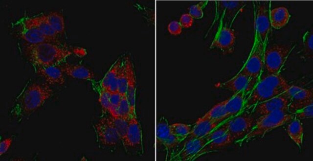
Immunocytochemistry Analysis: A 1:250 dilution from a representative lot detected laminin receptor in A431, HeLa, HepG2, and NIH/3T3 cells.

Biochem/physiol Actions

This polyclonal antibody detects the 37, 67, and 120 kDa forms of laminin receptor by targeting a sequence in the region between PPP1R16B-interacting and laminin-binding domains.

Physical form

Affinity purified.
Purified rabbit polyclonal antibody in buffer containing 0.1 M Tris-Glycine (pH 7.4), 150 mM NaCl with 0.05% sodium azide.

Preparation Note

Stable for 1 year at 2-8°C from date of receipt.

Analysis Note

Evaluated by Western Blotting in HeLa cell lysate.

Western Blotting Analysis: 1 µg/mL of this antibody detected laminin receptor in 10 µg of HeLa cell lysate.

Other Notes

Concentration: Please refer to lot specific datasheet.

Disclaimer

Unless otherwise stated in our catalog or other company documentation accompanying the product(s), our products are intended for research use only and are not to be used for any other purpose, which includes but is not limited to, unauthorized commercial uses, in vitro diagnostic uses, ex vivo or in vivo therapeutic uses or any type of consumption or application to humans or animals.
Ta strona może zawierać tekst przetłumaczony maszynowo.

Porównaj podobne pozycje

Wyświetl pełne porównanie

Pokaż różnice

1 of 1

Ta pozycja
L9393MABT1577MABT39
Quality Level

100

Quality Level

300

Quality Level

200

Quality Level

100

conjugate

unconjugated

conjugate

unconjugated

conjugate

unconjugated

conjugate

unconjugated

antibody form

affinity isolated antibody

antibody form

affinity isolated antibody

antibody form

purified antibody

antibody form

purified antibody

biological source

rabbit

biological source

rabbit

biological source

mouse

biological source

mouse

technique(s)

immunocytochemistry: suitable, western blot: suitable

technique(s)

dot blot: 1:1,000, immunohistochemistry (formalin-fixed, paraffin-embedded sections): 1:30 using human and animal tissues, microarray: suitable

technique(s)

immunocytochemistry: suitable, immunoprecipitation (IP): suitable, western blot: suitable

technique(s)

immunohistochemistry: suitable

clone

polyclonal

clone

polyclonal

clone

3E12, monoclonal

clone

4B12, monoclonal


Still not finding the right product?

Wypróbuj nasze narzędzie Narzędzie selektora produktów, aby zawęzić opcje.


Klasa składowania

12 - Non Combustible Liquids

wgk

WGK 1

flash_point_f

Not applicable

flash_point_c

Not applicable


Certyfikaty analizy (CoA)

Poszukaj Certyfikaty analizy (CoA), wpisując numer partii/serii produktów. Numery serii i partii można znaleźć na etykiecie produktu po słowach „seria” lub „partia”.

Masz już ten produkt?

Dokumenty związane z niedawno zakupionymi produktami zostały zamieszczone w Bibliotece dokumentów.

Odwiedź Bibliotekę dokumentów


Susanne Bürger et al.
International journal of molecular sciences, 22(1) (2021-01-06)
The demise of retinal ganglion cells (RGCs) is characteristic of diseases of the retina such as glaucoma and diabetic or ischemic retinopathies. Pigment epithelium-derived factor (PEDF) is a multifunctional secreted protein that mediates neuroprotection and inhibition of angiogenesis in the


Numer pozycji handlu globalnego

SKUNUMER GTIN
ABC93404055977353457

Questions

Reviews

No rating value

Active Filters