
MABE1007

Anti-TEL-AML1 fusion Antibody, clone 6F2
別名:
TEL-AML1 fusion|ETV6-RUNX1|Transcription factor ETV6|ETS translocation variant 6|ETS-related protein Tel1|Tel|Runt-related transcription factor 1|Acute myeloid leukemia 1 protein|Core-binding factor subunit alpha-2|CBF-alpha-2|Oncogene AML-1|Polyomavirus
サイズを選択してください
| 容量 | カタログ番号 | 在庫状況 | 単価 |
|---|
この商品について
biological source
rat
Quality Level
clone
monoclonal
species reactivity
human
technique(s)
ChIP: suitable, flow cytometry: suitable, western blot: suitable
General description
Immunogen
Application
Chromatin Immunoprecipitation Analysis
Analysis Note
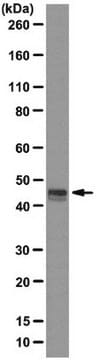
Western Blotting Analysis: A 1:20,000 dilution of this antibody detected TEL-AML1 fusion induction in 2.5 ug lysate from mifepristone-treated murine BA/F3 cells expressing inducible TEL-AML1 fusion.
Other Notes
1 of 1
当該品目 | |||
|---|---|---|---|
| clone monoclonal | clone R3-5G4, monoclonal | clone 3H3, monoclonal | clone polyclonal |
| biological source rat | biological source mouse | biological source mouse | biological source rabbit |
| species reactivity human | species reactivity human | species reactivity human | species reactivity human |
| technique(s) ChIP: suitable, western blot: suitable, flow cytometry: suitable | technique(s) immunoprecipitation (IP): suitable, western blot: suitable | technique(s) western blot: suitable | technique(s) ChIP: 1-10 μg (per reaction), immunoblotting: 0.04-0.4 μg/mL, immunofluorescence: 0.25-2 μg/mL, immunohistochemistry: 1:200-1:500 |
| Quality Level 300 | Quality Level 100 | Quality Level 100 | Quality Level 100 |


試験成績書(COA)
製品のロット番号・バッチ番号を入力して、試験成績書(COA) を検索できます。ロット番号・バッチ番号は、製品ラベルに「Lot」または「Batch」に続いて記載されています。
関連コンテンツ
A major focus of breast cancer research is to understand the mechanisms responsible for disease progression and drug resistance. Toward that end, it has been found that approximately two thirds of all human breast carcinomas overexpress the Estrogen Receptor α (ERα) protein and it remains the primary pharmacological target for endocrine therapy1,2. The normal cellular function of ERα is as a transcription factor that mediates a wide variety of physiological processes, many of which are dependent upon phosphorylation of the receptor at specific amino acid residues3,4. Indeed, ERα is known to be phosphorylated at a multitude of different sites, yet how these all correlate to disease remains unclear5. Here, we interrogated multiple sites of ERα for phosphorylation status by screening an extensive panel of different breast cancer patient samples and other non-breast cancer tissue microarray (TMA) slide samples to determine their relevance to disease.
Historically chromatin was considered to contain only DNA, histones, non-histone proteins and transiently associated mRNAs. While this remains true, current data suggest that a variety of non-coding RNAs (e.g. long non-coding RNAs, enhancer RNAs and even miRNAs) also associate with chromatin and serve regulatory functions.
グローバルトレードアイテム番号
| カタログ番号 | GTIN |
|---|---|
| MABE1007 | 04055977168259 |