イメージング解析・ライブセルイメージング

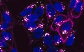
ライブセルのイメージングと解析により、科学者は動的な細胞事象をリアルタイムで調べて、生物学に対する独自の洞察を得ることができます。これらの手法は、PCRや、細胞事象をスナップショットとして解析するフローサイトメトリー、免疫細胞染色、抗体による組織染色とは対照的です。今日、カメラやライブセル蛍光色素、蛍光タンパク質、動画圧縮、タイムラプス顕微鏡観察技術の向上により、ライブセル、細胞間相互作用、および細胞内プロセスを驚くほど詳細かつ忠実に視覚化し、解析できるようになっています。
注目のカテゴリー
CellASIC® ONIX2 Microfluidic Systemは、哺乳類、酵母、および細菌細胞のライブセルイメージングおよび解析のための細胞培養環境の正確な操作を可能にする、パワフルな自動プラットフォームです。
ライブセルイメージングシステム
細胞の複雑な構造や挙動は、さまざまな手法で視覚化できます。静的解析では、細胞を固定処理して染色しますが、細胞を死滅させて、その時点における細胞状態をとらえることが必要となります。静的手法は一部の用途には適していますが、細胞活性に関する洞察を提供するものではありません。動的なライブセルアプローチでは、従来の染色なしで、ライブセルの構造、挙動、または組成を容易に視覚化できます。これらの動的手法は、in vivo条件を模倣する必要のある用途や、薬物スクリーニングの予測アッセイの開発に最適です。
ライブセルイメージングシステムでは、哺乳類、細菌、および酵母細胞の長期研究を可能にする、高度な光学機能と環境制御、および各種の試薬が使用されます。マイクロ流体を採用した培養およびイメージングシステムにより、細胞への物理的ストレスを制限しながら、流量、気体混合物、温度などの培養微小環境を正確に制御できます。ライブセルのイメージングと解析は、低酸素症、細胞遊走、3D細胞培養、細胞骨格動態、タンパク質輸送など、さまざまな用途に適しています。高度なシステムは、培養条件、処理の管理と中止、イメージング間隔を自動化して、静止画または動画データとして表示される画像を作成します。
ライブセルイメージング試薬
ライブセルイメージング用の培地とサプリメントは、タイムラプス実験中に光による細胞損傷から細胞を保護するように設計されています。これらの試薬はまた、自家蛍光と光退色を抑制するように処方されており、ライブセル蛍光イメージングの画質を劇的に向上させます。
ライブセルの動的プロセスを研究するための、さまざまなライブセル染色色素が提供されています。ライブセル蛍光オルガネラ色素は、細胞膜、核、細胞質、ミトコンドリア、リソソーム、小胞体(ER)、ゴルジ体、細胞骨格タンパク質などの特定のオルガネラを、細胞毒性を増加させることなく選択的に染色することができます。ライブセルイメージングの対比染色としてのオルガネラ色素は、機能研究にも役立てられています。
GFPとRFPを備えたバイオセンサーを使用すると、特定のタンパク質とライブセル内のそのタンパク質の細胞内位置を、蛍光顕微鏡またはタイムラプス動画キャプチャのいずれかによって検出できます。レンチウイルスベクターシステムは、細胞内の遺伝子や遺伝子産物の導入によく使われるツールであり、化学物質ベースのトランスフェクションに勝る多くの利点があります。細胞透過性のライブセル色素は、細胞小器官の検出のほかに、アポトーシスの検出や、細胞生存率、細胞健全性、低酸素症、活性酸素種の研究、カルシウムインジケーターイメージング、神経細胞または幹細胞実験などの用途にも利用されています。
関連資料
- Cell based assays for cell proliferation (BrdU, MTT, WST1), cell viability and cytotoxicity experiments for applications in cancer, neuroscience and stem cell research.
- Firefly luciferase is a sensitive reporter for gene studies due to its absence in mammalian cells or tissues.
- PKH and CellVue® Fluorescent Cell Linker Kits provide fluorescent labeling of live cells over an extended period of time, with no apparent toxic effects.
- Fluorescent calcium indicators and dyes to measure Ca+ flux used in calcium imaging experiments. Calcium ions (Ca2+) play vital cellular physiology roles in signal transduction pathways, in neurotransmitter release, in contraction of all muscle cell types, as enzyme cofactors, and in fertilization.
- Lipophilic cell tracking dyes enable cancer biologists to track tumor and immune cell functions both in vitro and in vivo. Read the article to choose a right membrane dye kit for cell tracking and proliferation monitoring.
- すべて表示 (35)
関連プロトコル
- PKH dyes label exosomes for tracking experiments in vitro and in vivo, detailed protocol provided.
- Cell staining can be divided into four steps: cell preparation, fixation, application of antibody, and evaluation.
- ICC Cell Capture Imaging Reagent simplifies imaging, ideal for rare cell and low cell count samples.
- Toluidine blue selectively stains nuclear material and acidic tissue components, aiding in histological staining for tissues rich in DNA/RNA.
- This protocol covers 3 modes for the microscopic examination of cell samples.
- すべて表示 (6)
技術資料・プロトコルの検索
お問い合わせ
ご不明な点がございましたら、お問い合わせページをご覧いただくか
、テクニカルサービスまでお問い合わせください。
メール:[email protected]
電話:03-6756-8245
サポート
- Chromatogram Search
Use the Chromatogram Search to identify unknown compounds in your sample.
- 計算ツール・アプリ
ウェブツールボックス-分析化学、ライフサイエンス、化学合成および材料科学のためのサイエンスリサーチツールとリソース
- Customer Support Request
お問い合わせページでは、製品のお問い合わせや、注文や納期、お取引やアカウント、ウェブサイトに関するお問い合わせをお申込みいただけます。
- FAQ
Explore our Frequently Asked Questions for answers to commonly asked questions about our products and services.
続きを確認するには、ログインするか、新規登録が必要です。
アカウントをお持ちではありませんか?


