
05-765
Anti-MLL/HRX-Antikörper, CT, Klon 9-12
clone 9-12, Upstate®, from mouse
Synonym(e):
Histone-lysine N-methyltransferase 2A, Lysine N-methyltransferase 2A, ALL-1, CXXC-type zinc finger protein 7, Myeloid/lymphoid or mixed-lineage leukemia, Myeloid/lymphoid or mixed-lineage leukemia protein 1, Trithorax-like protein, Zinc finger protein HR
Größe auswählen
| Packungsgröße | SKU | Verfügbarkeit | Preis |
|---|---|---|---|
| 200 μg | Warenkorb auf Verfügbarkeit prüfen | € 583,00 |
Über diesen Artikel
biological source
mouse
Quality Level
conjugate
unconjugated
antibody form
purified immunoglobulin
antibody product type
primary antibodies
clone
9-12, monoclonal
species reactivity
mouse, human
manufacturer/tradename
Upstate®
technique(s)
ChIP: suitable, immunofluorescence: suitable, immunoprecipitation (IP): suitable, western blot: suitable
isotype
IgG1
NCBI accession no.
UniProt accession no.
shipped in
wet ice
target post-translational modification
unmodified
Gene Information
human ... KMT2A(4297)
General description
Immunogen
Application
Epigenetik & Zellkernfunktion
Histone
Chromatin-Immunpräzipitation(ChIP)-Analyse: Eine repräsentative Charge wies die MLL-Belegung an Hoxa10 und Meis-Promotoren in Maus-MLL-AF10-Leukämiezellen nach (Gallo, M., et al. (2013). Cancer Res. 73(1):417-427).
Chromatin-Immunpräzipitation(ChIP)-Analyse: Eine repräsentative Charge wies eine verstärkte MLL-Belegung an am Ink4a-Locus von 8 Monate alten im Vergleich zu 2 Monate alten bEzTG-Maus-Inselzellen nach. Dieselbe altersabhängige Ink4a-Locus-Anreicherung wurde in H3K4me3 festgestellt, während der gegenläufige Trend bei der Ezh- und H3K27me3-Anreicherung am selben Locus festgestellt wurde (Zhou, J.X., et al. (2013). J. Clin. Invest. 123(11):4849-4858).
Chromatin-Immunpräzipitation (ChIP)-Analyse: Eine repräsentative Charge wies die MLL-Belegung in der HOXA10-Promotor-Region in humanen neuralen G179NS- und G411NS-Glioblastom-Stammzellen nach (Gallo, M., et al. (2013). Cancer Res. 73(1):417-427).
Chromatin-Immunpräzipitation(ChIP)-Analyse: Eine repräsentative Charge wies die MLL-Belegung am DNA-Replikationsursprung (RD) sowie am Exon 1b und am gemeinsamen Exon 2 von p16INK4a/p19ARF in embryonalen Mausfibroblasten (MEF) nach. In seneszenten und Polycomb-mutanten MEF wurde an diesen Stellen eine gesteigerte MLL-Anreicherung festgestellt (Agherbi, H., et al. (2009). PLoS One. 4(5):e5622).
Chromatin-Immunpräzipitation (ChIP)-Analyse: Eine repräsentative Charge wies die MLL-Belegung in der Hoxa9-AB-Region mit embryonalen Mausfibroblasten(MEF)-Chromatinaufbereitungen nach (Erfurth, F. E., et al. (2008). Proc. Natl. Acad. Sci. U.S.A. 105(21):7517-7522).
Immunpräzipitations-Analyse: Eine repräsentative Charge co-immunpräzipitierte JmjD3 und RbBP5, aber nicht Dnmt3a mit MLL aus Min6-Mausinsulinomzellextrakt (Zhou, J.X., et al. (2013). J. Clin. Invest. 123(11):4849-4858).
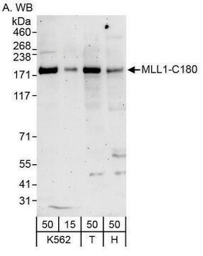
Western-Blot-Analyse: Eine repräsentative Charge wies höhere MLL-Werte in kultivierten neuralen Glioblastomstammzellen (GNS) als in neuralen Stammzellen (NS) sowie eine MLL-Anreicherung in der CD15+-Fraktion von frisch reserzierten Glioblastomzellen (GBM) nach (Gallo, M., et al. (2013). Cancer Res. 73(1):417-427).
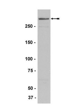
Western-Blot-Analyse: Eine repräsentative Charge wies das SET-Domäne-haltige C-terminale Fragment der enzymatischen MLL-Komplex-Untereinheit MLL (C180; MLLC) in Anti-FLAG-Immunpräzipitat aus HeLaS-Zellen, die FLAG-markiertes hDPY-30 stabil exprimieren, nach (Cho, Y.W., et al. (2007). J. Biol. Chem. 282(28): 20395-20406).
Biochem/physiol Actions
Physical form
Preparation Note
Analysis Note
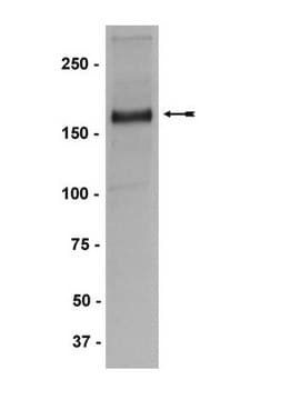
K562-Zellkernextrakt
Western-Blot-Analyse: 0,1–1 µg/ml dieses Antikörpers wies MLL-C-terminales Fragment (C180; MLLC) in K562-Zellkernextrakt nach.
Other Notes
Legal Information
Disclaimer
1 of 1
Dieser Artikel | |||
|---|---|---|---|
| antibody form purified immunoglobulin | antibody form affinity purified immunoglobulin | antibody form affinity purified immunoglobulin | antibody form affinity isolated antibody |
| species reactivity mouse, human | species reactivity mouse, human | species reactivity human | species reactivity rat, human, mouse |
| biological source mouse | biological source mouse | biological source rabbit | biological source rabbit |
| clone 9-12, monoclonal | clone N4.4, monoclonal | clone polyclonal | clone polyclonal |
| conjugate unconjugated | conjugate unconjugated | conjugate unconjugated | conjugate unconjugated |
| Gene Information human ... KMT2A(4297) | Gene Information human ... KMT2A(4297) | Gene Information rabbit ... MLL1(4297) | Gene Information human ... KMT2C(58508) |
Still not finding the right product?
Probieren Sie unser Produkt-Auswahlhilfe, um Ihre Auswahl einzugrenzen
Lagerklasse
10 - Combustible liquids
wgk
WGK 1
Analysenzertifikate (COA)
Suchen Sie nach Analysenzertifikate (COA), indem Sie die Lot-/Chargennummer des Produkts eingeben. Lot- und Chargennummern sind auf dem Produktetikett hinter den Wörtern ‘Lot’ oder ‘Batch’ (Lot oder Charge) zu finden.
Besitzen Sie dieses Produkt bereits?
In der Dokumentenbibliothek finden Sie die Dokumentation zu den Produkten, die Sie kürzlich erworben haben.
Verwandter Inhalt
Cancer is a complex disease manifestation. At its core, it remains a disease of abnormal cellular proliferation and inappropriate gene expression. In the early days, carcinogenesis was viewed simply as resulting from a collection of genetic mutations that altered the gene expression of key oncogenic genes or tumor suppressor genes leading to uncontrolled growth and disease (Virani, S et al 2012). Today, however, research is showing that carcinogenesis results from the successive accumulation of heritable genetic and epigenetic changes. Moreover, the success in how we predict, treat and overcome cancer will likely involve not only understanding the consequences of direct genetic changes that can cause cancer, but also how the epigenetic and environmental changes cause cancer (Johnson C et al 2015; Waldmann T et al 2013). Epigenetics is the study of heritable gene expression as it relates to changes in DNA structure that are not tied to changes in DNA sequence but, instead, are tied to how the nucleic acid material is read or processed via the myriad of protein-protein, protein-nucleic acid, and nucleic acid-nucleic acid interactions that ultimately manifest themselves into a specific expression phenotype (Ngai SC et al 2012, Johnson C et al 2015). This review will discuss some of the principal aspects of epigenetic research and how they relate to our current understanding of carcinogenesis. Because epigenetics affects phenotype and changes in epigenetics are thought to be key to environmental adaptability and thus may in fact be reversed or manipulated, understanding the integration of experimental and epidemiologic science surrounding cancer and its many manifestations should lead to more effective cancer prognostics as well as treatments (Virani S et al 2012).