
ABN76
Anti-Neurofilament H (200 kDa)-Antikörper
rabbit polyclonal
Synonym(e):
Neurofilament heavy polypeptide, Neurofilament H
Größe auswählen
| Packungsgröße | SKU | Verfügbarkeit | Preis |
|---|---|---|---|
| 100 μg | Warenkorb auf Verfügbarkeit prüfen | € 401,00 |
Über diesen Artikel
€ 401,00
Produktname
Anti-Neurofilament H (200 kDa)-Antikörper, from rabbit, purified by affinity chromatography
biological source
rabbit
Quality Level
antibody form
affinity isolated antibody
antibody product type
primary antibodies
clone
polyclonal
purified by
affinity chromatography
species reactivity
mouse, human, rat, bovine
technique(s)
immunohistochemistry: suitable (paraffin), western blot: suitable
NCBI accession no.
UniProt accession no.
shipped in
wet ice
target post-translational modification
unmodified
Gene Information
bovine ... Nefh(528842)
human ... NEFH(4744)
mouse ... Nefh(380684)
rat ... Nefh(24587)
General description
Immunogen
Application
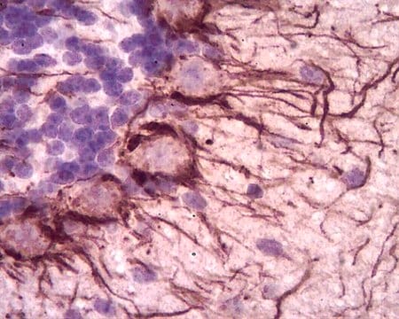
Analysis Note
Western-Blot-Analyse: Mit 0,1 µg/ml dieses Antikörpers wurde Neurofilament H in Gewebelysat aus dem bovinen Cerebellum nachgewiesen.
Bovines Cerebellum-Gewebelysat
Other Notes
1 of 1
Dieser Artikel | |||
|---|---|---|---|
| Quality Level 100 | Quality Level 100, 300 | Quality Level 100 | Quality Level 100 |
| antibody form affinity isolated antibody | antibody form purified immunoglobulin | antibody form serum | antibody form serum |
| biological source rabbit | biological source mouse | biological source rabbit | biological source rabbit |
| technique(s) immunohistochemistry: suitable (paraffin), western blot: suitable | technique(s) immunohistochemistry: suitable, western blot: suitable | technique(s) immunohistochemistry: suitable, western blot: suitable | technique(s) immunohistochemistry (formalin-fixed, paraffin-embedded sections): suitable, western blot: suitable |
| Gene Information bovine ... Nefh(528842) | Gene Information human ... NEFH(4744) | Gene Information bovine ... Nefh(528842) | Gene Information human ... NEFH(4744) |
| shipped in wet ice | shipped in wet ice | shipped in dry ice | shipped in dry ice |


Still not finding the right product?
Explore all of our products under Anti-Neurofilament H (200 kDa)-Antikörper
— oder —
Probieren Sie unser Produkt-Auswahlhilfe, um Ihre Auswahl einzugrenzen
Lagerklasse
12 - Non Combustible Liquids
wgk
WGK 1
flash_point_f
Not applicable
flash_point_c
Not applicable
Analysenzertifikate (COA)
Suchen Sie nach Analysenzertifikate (COA), indem Sie die Lot-/Chargennummer des Produkts eingeben. Lot- und Chargennummern sind auf dem Produktetikett hinter den Wörtern ‘Lot’ oder ‘Batch’ (Lot oder Charge) zu finden.
Besitzen Sie dieses Produkt bereits?
In der Dokumentenbibliothek finden Sie die Dokumentation zu den Produkten, die Sie kürzlich erworben haben.
Verwandter Inhalt
Global Trade Item Number
| SKU | GTIN |
|---|---|
| ABN76 | 04053252632051 |