Direkt zum Inhalt
Merck

Fortfahren mit

P8333

Anti-Protein Kinase Cδ antibody produced in rabbit

whole antiserum

Synonym(e):

Anti-PKC δ

Anmelden zur Ansicht der Organisations- und Vertragspreise.

Größe auswählen

Ansicht ändern
PackungsgrößeSKUVerfügbarkeitPreis
0.2 mL
Warenkorb auf Verfügbarkeit prüfen
€ 361,00

Über diesen Artikel

UNSPSC Code:
12352203
NACRES:
NA.44
MDL number:
Conjugate:
unconjugated
Clone:
polyclonal
Application:
ARR, DB, WB
Citations:
10

€ 361,00


Warenkorb auf Verfügbarkeit prüfen

Großbestellung anfragen
Technischer Dienst
Benötigen Sie Hilfe? Unser Team von erfahrenen Wissenschaftlern ist für Sie da.
Unterstützung erhalten


biological source

rabbit

Quality Level

conjugate

unconjugated

antibody form

whole antiserum

antibody product type

primary antibodies

clone

polyclonal

contains

15 mM sodium azide

species reactivity

rat

technique(s)

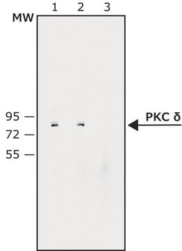
dot blot: 1:50,000, microarray: suitable, western blot: 1:10,000 using rat brain extract

UniProt accession no.

shipped in

dry ice

storage temp.

−20°C

target post-translational modification

unmodified

Gene Information

rat ... Prkcd(170538)

General description

Protein Kinase C (PKC) is a family of serine/threonine (Ser/Thr)-specific protein kinases. PKC is a phospholipid-dependent enzyme, activated by the lipid 1,2-diacylglycerol (DAG). The protein kinase C delta (PKC δ) isoenzyme appears to be widely expressed in the brain, lung, heart, spleen, liver, ovary, pancreas, thymus, adrenal gland, skin and rat embryonic fibroblasts, and is expressed in lower levels in certain mouse fibroblasts. PKD is also located in the cytosol, nuclear compartment and in mitochondria in response to cellular stress.

Immunogen

Synthetic peptide corresponding to the C-terminal variable (V5) region (amino acids 662-673) of rat PKC δ coupled to KLH with glutaraldehyde.

Application

Anti- protein kinase c δ antibody may be used in:
  • immunoprecipitation
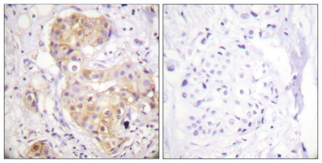
  • immunohistochemistry
  • immunoblotting
  • ELISA
  • chemiluminescence detection systems to detect PKC δ
  • dot-blot immunoassay
Applications in which this antibody has been used successfully, and the associated peer-reviewed papers, are given below.
Western Blotting (1 paper)

Biochem/physiol Actions

Anti-Protein Kinase C δ specifically reacts in dot-blot immunoassay with PKC δ peptide conjugated to BSA with 1-ethyl-3-(3-dimethylamino-propyl)-carbodiimide (EDCI).
Protein Kinase C isotype δ (PKCδ) modulates the inflammatory response. It acts as a signal transducer of several signaling pathways. Hence it can be considered as a vital therapeutic target to treat sepsis induced-lung injury. In sepsis, PKCδ participates in platelet-mediated activation. Overexpression and stimulation of PKC δ leads to cell division arrest in Chinese hamster ovary (CHO) cells and growth inhibition of NIH3T3 cells.
Protein kinase C (PKC) has a pivotal role in cell growth and differentiation, modulation of neurotransmission, signal transduction and oncogenesis. Anti-protein kinase c δ antibody can be used for studying the differential tissue expression and intracellular localization of PKC δ. It can also be used in western blotting and microarray.

Physical form

Rabbit Anti-Protein Kinase C δ is supplied as liquid containing 0.1% sodium azide as preservative.

Preparation Note

For continuous use, store at 2-8 °C for up to one month. For extended storage freeze in working aliquots. Repeated freezing and thawing is not recommended.Storage in "frost-free" freezers is not recommended. If slight turbidity occurs upon prolonged storage, clarify the solution by centrifugation before use.

Disclaimer

Unless otherwise stated in our catalog or other company documentation accompanying the product(s), our products are intended for research use only and are not to be used for any other purpose, which includes but is not limited to, unauthorized commercial uses, in vitro diagnostic uses, ex vivo or in vivo therapeutic uses or any type of consumption or application to humans or animals.

Ähnliche Artikel vergleichen

Vollständigen Vergleich anzeigen

Unterschiede anzeigen

1 of 1

Dieser Artikel
P5704SAB4502360SAB1412060
antibody form

whole antiserum

antibody form

ascites fluid

antibody form

affinity isolated antibody

antibody form

purified immunoglobulin

biological source

rabbit

biological source

mouse

biological source

rabbit

biological source

mouse

conjugate

unconjugated

conjugate

unconjugated

conjugate

unconjugated

conjugate

unconjugated

Quality Level

200

Quality Level

200

Quality Level

100

Quality Level

-

clone

polyclonal

clone

MC5, monoclonal

clone

polyclonal

clone

8H6, monoclonal

technique(s)

dot blot: 1:50,000, western blot: 1:10,000 using rat brain extract, microarray: suitable

technique(s)

immunohistochemistry (frozen sections): suitable, immunoprecipitation (IP): suitable, microarray: suitable, western blot: 1:500 using rat brain cytosol preparation

technique(s)

ELISA: 1:1000, immunohistochemistry: 1:50-1:100, western blot: 1:500-1:1000

technique(s)

indirect ELISA: suitable, western blot: 1-5 μg/mL


Still not finding the right product?

Probieren Sie unser Produkt-Auswahlhilfe, um Ihre Auswahl einzugrenzen


Hier finden Sie alle aktuellen Versionen:

Analysenzertifikate (COA)

Lot/Batch Number

Die passende Version wird nicht angezeigt?

Wenn Sie eine bestimmte Version benötigen, können Sie anhand der Lot- oder Chargennummer nach einem spezifischen Zertifikat suchen.

Besitzen Sie dieses Produkt bereits?

In der Dokumentenbibliothek finden Sie die Dokumentation zu den Produkten, die Sie kürzlich erworben haben.

Die Dokumentenbibliothek aufrufen


Verwandter Inhalt


Ikuko Koyama-Honda et al.
Autophagy, 9(10), 1491-1499 (2013-07-26)
Autophagosome formation is governed by sequential functions of autophagy-related (ATG) proteins. Although their genetic hierarchy in terms of localization to the autophagosome formation site has been determined, their temporal relationships remain largely unknown. In this study, we comprehensively analyzed the
Protein kinase D activation induces mitochondrial fragmentation and dysfunction in cardiomyocytes
Jhun BS, et al.
The Journal of Physiology, 596(5), 827-855 (2018)
Junping Zhang et al.
Molecular therapy. Methods & clinical development, 32(1), 101215-101215 (2024-03-11)
The unique palindromic inverted terminal repeats (ITRs) and single-stranded nature of adeno-associated virus (AAV) DNA are major hurdles to current sequencing technologies. Due to these characteristics, sequencing noncanonical AAV genomes present in AAV vector preparations remains challenging. To address this


Global Trade Item Number

SKUGTIN
P8333-.2ML04061837914201

Questions

Reviews

No rating value

Active Filters