
ABS1511

Anti-Neph1 Antibody, cytoplasmic domain
from rabbit, purified by affinity chromatography
Synonym(e):
Kin of IRRE-like protein 1, Kin of irregular chiasm-like protein 1, Nephrin-like protein 1, Neph1, cytoplasmic domain
Größe auswählen
| Packungsgröße | SKU | Verfügbarkeit | Preis |
|---|
Über diesen Artikel
biological source
rabbit
Quality Segment
conjugate
unconjugated
antibody form
affinity isolated antibody
antibody product type
primary antibodies
clone
polyclonal
purified by
affinity chromatography
species reactivity
mouse, rat, human
technique(s)
electron microscopy: suitable, immunocytochemistry: suitable, immunofluorescence: suitable, immunoprecipitation (IP): suitable, western blot: suitable
NCBI accession no.
UniProt accession no.
shipped in
wet ice
target post-translational modification
unmodified
Gene Information
human ... KIRREL1(55243)
General description
Immunogen
Application
Signaling
Signaling Neuroscience
Western Blotting Analysis: A representative lot detected ischemia-induced Neph1 membrane-to-cytosol translocation in human podocytes (Wagner, M.C., et al. (2008). J Biol Chem. 283(51):35579-35589).
Western Blotting Analysis: A representative lot detected Neph1 in mouse glomeruli & cultured human podocytes (Arif, E., et al. (2011). Mol Cell Biol. 31(10):2134-2150).
Immunoprecipitation Analysis: A representative lot immunoprecipitated Neph1 from rat glomerular and human podocyte cell lysates (Arif, E., et al. (2011). Mol Cell Biol. 31(10):2134-2150).
Immunofluorescence Analysis: A representative lot detected Neph1 using both paraffin-embedded and frozen rat kidney sections (Arif, E., et al. (2011). Mol Cell Biol. 31(10):2134-2150; Barletta, G.M., et al. (2003). J Biol Chem. 278(21):19266-19271).
Electron Microscopy Analysis: A representative lot detected Neph1 in frozen rat kidney sections (Barletta, G.M., et al. (2003). J Biol Chem. 278(21):19266-19271).
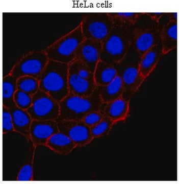
Immunocytochemistry Analysis: A representative lot detected Neph1 in cultured human podocytes (Arif, E., et al. (2014). J Biol Chem. 289(14):9502-9518; Arif, E., et al. (2011). Mol Cell Biol. 31(10):2134-2150; Wagner, M.C., et al. (2008). J Biol Chem. 283(51):35579-35589).
Biochem/physiol Actions
Physical form
Preparation Note
Handling Recommendations: Upon receipt and prior to removing the cap, centrifuge the vial and gently mix the solution. Aliquot into microcentrifuge tubes and store at -20°C. Avoid repeated freeze/thaw cycles, which may damage IgG and affect product performance.
Note: Variability in freezer temperatures below -20°C may cause glycerol containing solutions to become frozen during storage.
Analysis Note
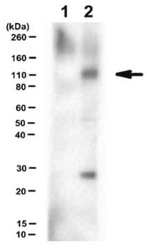
Western Blotting Analysis: A 1:1,000 dilution of this antibody detected Neph1 in rat kidney tissue lysate.
Other Notes
Disclaimer
1 of 1
Dieser Artikel | |||
|---|---|---|---|
| Quality Level 100 | Quality Level 100 | Quality Level 100 | Quality Level 100 |
| biological source rabbit | biological source rabbit | biological source mouse | biological source rabbit |
| antibody form affinity isolated antibody | antibody form affinity isolated antibody | antibody form affinity isolated antibody | antibody form serum |
| conjugate unconjugated | conjugate - | conjugate unconjugated | conjugate unconjugated |
| UniProt accession no. | UniProt accession no. | UniProt accession no. | UniProt accession no. |
| Gene Information human ... KIRREL1(55243) | Gene Information human ... KIRREL1(55243) | Gene Information human ... KRIT1(889) | Gene Information human ... SMARCAL1(50485) |

Still not finding the right product?
Probieren Sie unser Produkt-Auswahlhilfe, um Ihre Auswahl einzugrenzen
Lagerklasse
10 - Combustible liquids
wgk
WGK 3
Analysenzertifikate (COA)
Suchen Sie nach Analysenzertifikate (COA), indem Sie die Lot-/Chargennummer des Produkts eingeben. Lot- und Chargennummern sind auf dem Produktetikett hinter den Wörtern ‘Lot’ oder ‘Batch’ (Lot oder Charge) zu finden.
Besitzen Sie dieses Produkt bereits?
In der Dokumentenbibliothek finden Sie die Dokumentation zu den Produkten, die Sie kürzlich erworben haben.
Verwandter Inhalt
A major focus of breast cancer research is to understand the mechanisms responsible for disease progression and drug resistance. Toward that end, it has been found that approximately two thirds of all human breast carcinomas overexpress the Estrogen Receptor α (ERα) protein and it remains the primary pharmacological target for endocrine therapy1,2. The normal cellular function of ERα is as a transcription factor that mediates a wide variety of physiological processes, many of which are dependent upon phosphorylation of the receptor at specific amino acid residues3,4. Indeed, ERα is known to be phosphorylated at a multitude of different sites, yet how these all correlate to disease remains unclear5. Here, we interrogated multiple sites of ERα for phosphorylation status by screening an extensive panel of different breast cancer patient samples and other non-breast cancer tissue microarray (TMA) slide samples to determine their relevance to disease.
Global Trade Item Number
| SKU | GTIN |
|---|---|
| ABS1511 | 04055977176544 |