
09-403
Anti-TKS5(SH3 #1)-Antikörper
from rabbit
Synonym(e):
Adaptor protein TKS5, Five SH3 domain-containing protein, SH3 and PX domains 2A, SH3 multiple domains 1, SH3 multiple domains protein 1, five SH3 domains
Größe auswählen
| Packungsgröße | SKU | Verfügbarkeit | Preis |
|---|---|---|---|
| 100 μg | Warenkorb auf Verfügbarkeit prüfen | € 478,00 |
Über diesen Artikel
biological source
rabbit
Quality Level
conjugate
unconjugated
antibody form
purified antibody
antibody product type
primary antibodies
clone
polyclonal
species reactivity
mouse, human
technique(s)
immunocytochemistry: suitable, immunoprecipitation (IP): suitable, western blot: suitable
NCBI accession no.
UniProt accession no.
shipped in
wet ice
target post-translational modification
unmodified
Gene Information
human ... SH3PXD2A(9644)
mouse ... Sh3Pxd2A(14218)
General description
Immunogen
Application
Zytoskelettale Signalisierung
Zellstruktur
Biochem/physiol Actions
Physical form
Preparation Note
Analysis Note
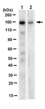
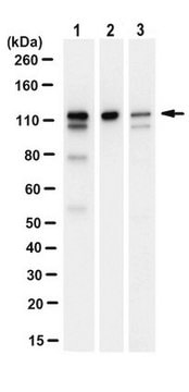
Western Blot:
NIH/3T3-Lysat (100 % Konfluenz)
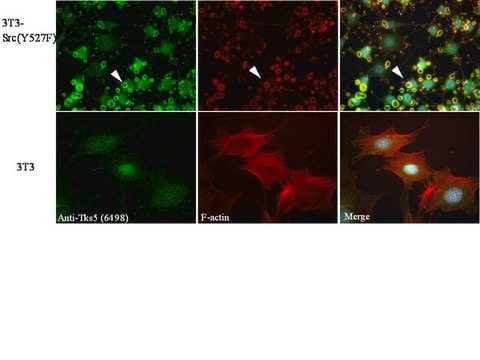
Immunzytochemie:
Maus-3T3-Src(Y527F)-Zellen
Other Notes
Disclaimer
1 of 1
Dieser Artikel | |||
|---|---|---|---|
| species reactivity mouse, human | species reactivity mouse, human | species reactivity human | species reactivity human |
| clone polyclonal | clone polyclonal | clone 13H6.3, monoclonal | clone 9H2.1, monoclonal |
| conjugate unconjugated | conjugate unconjugated | conjugate unconjugated | conjugate unconjugated |
| antibody form purified antibody | antibody form purified antibody | antibody form purified antibody | antibody form purified immunoglobulin |
| biological source rabbit | biological source rabbit | biological source mouse | biological source mouse |
| shipped in wet ice | shipped in wet ice | shipped in ambient | shipped in ambient |
Still not finding the right product?
Probieren Sie unser Produkt-Auswahlhilfe, um Ihre Auswahl einzugrenzen
Lagerklasse
12 - Non Combustible Liquids
wgk
WGK 1
flash_point_f
Not applicable
flash_point_c
Not applicable
Analysenzertifikate (COA)
Suchen Sie nach Analysenzertifikate (COA), indem Sie die Lot-/Chargennummer des Produkts eingeben. Lot- und Chargennummern sind auf dem Produktetikett hinter den Wörtern ‘Lot’ oder ‘Batch’ (Lot oder Charge) zu finden.
Besitzen Sie dieses Produkt bereits?
In der Dokumentenbibliothek finden Sie die Dokumentation zu den Produkten, die Sie kürzlich erworben haben.
Verwandter Inhalt
QCM™ Gelatin Invadopodia Assays provide optimized materials and protocols to enable reproducible analysis of invadopodia in invasive tumor cells (Catalog No. ECM670 for green fluorescence, Catalogue No. ECM671 for red fluorescence). Reagents are provided for coating glass culture surfaces with fluorescent matrix and for colocalizing the actin cytoskeleton and nuclei with invadopodial degradation sites. This assay may also be used for assessing the activity of inhibitors and promoters of invadopodia formation and function. Furthermore, different cell types and individual cells in heterogeneous populations may be analyzed for invasive potential. Finally, the assay kits provide troubleshooting suggestions, recommendations for coating on multiple substrate formats, and example studies in several assay systems (e.g., various cell types, time-course studies, degradation modulation).
Global Trade Item Number
| SKU | GTIN |
|---|---|
| 09-403 | 04053252620867 |