Accéder au contenu
MilliporeSigma

Passer à

D8043

Anti-Dystrophin Antibody

mouse monoclonal, MANDRA1

Se connecter pour consulter les tarifs organisationnels et contractuels.

Sélectionner une taille de conditionnement

Changer de vue
ConditionnementRéférenceDisponibilitéPrix
0.2 mL
Veuillez contacter notre Service Clients pour connaître la disponibilité de ce produit.
778,00 $

A propos de cet article

UNSPSC Code:
12352203
NACRES:
NA.41
MDL number:
Conjugate:
unconjugated
Clone:
MANDRA1, monoclonal
Application:
ARR, IF
Citations:
29

778,00 $


Veuillez contacter notre Service Clients pour connaître la disponibilité de ce produit.
Un anticorps recombinant, sans conservateur, est disponible pour votre cible. Essayez ZMS1051

Demander une commande groupée
Service technique
Besoin d'aide ? Notre équipe de scientifiques expérimentés est là pour vous.
Laissez-nous vous aider


Nom du produit

Monoclonal Anti-Dystrophin antibody produced in mouse, clone MANDRA1, ascites fluid

biological source

mouse

Quality Level

conjugate

unconjugated

antibody form

ascites fluid

antibody product type

primary antibodies

clone

MANDRA1, monoclonal

contains

15 mM sodium azide

species reactivity

rat, human, mouse, fish

technique(s)

indirect immunofluorescence: 1:100 using freshly dissected and frozen human or animal muscle tissue., microarray: suitable

isotype

IgG1

UniProt accession no.

shipped in

dry ice

storage temp.

−20°C

target post-translational modification

unmodified

Gene Information

human ... DMD(1756)
mouse ... Dmd(13405)
rat ... Dmd(24907)

General description

Dystrophin is a muscle membrane protein (427 kDa) which is absent, reduced or altered as a result of mutation in Duchenne and Becker muscular dystrophies (DMD/BMD) or its homologue in the mouse. Severe DMD is associated with a marked dystrophin deficiency whereas patients with the milder form of DMD show less pronounced abnormalities of protein expression. Because abnormalities in the protein expression occur specifically in patients with these types of muscular dystrophy, dystrophin analysis may be used to distinguish these conditions from other neuromuscular diseases. Predictions from the sequence suggest a structural protein on the inner face of the membrane, consisting of a 25-repeat, rod-like triple-helical domain separating an N-terminal actin-binding domain from two C-terminal domains, one of which is rich in cysteine. The large size of dystrophin and its low abundance (<0.01% of the total muscle protein) are a hindrance to the isolation of intact, native protein for structure/function studies.
The C-terminal domain of the human dystrophin molecule (amino acids residues 3558-3684) is present in normal muscle tissue. It is also present in nearly all Becker muscular dystrophies, but is absent in cases of Duchenne muscular dystrophies and in the dystrophic mouse (mdx).
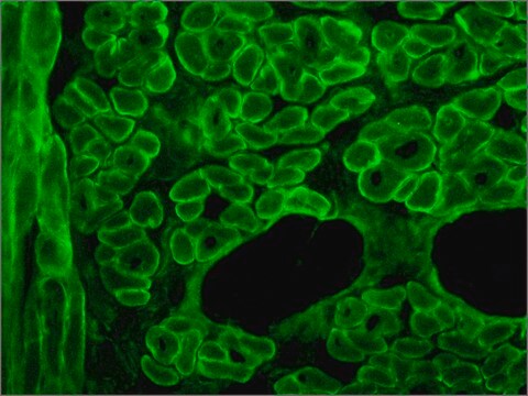
The antibody recognizes an epitope located on the 128 amino acids at the end of the C-terminal domain of the human dystrophin molecule (amino acid residues 3558-3684). Immunohistochemical staining of muscle tissue results in a clear labeling confined to the periphery (plasma membrane) of normal striated muscle fibers. By immunoblotting, the antibody stains dystrophin (427 kDa) in muscle and brain extracts. It also stains the 70-75 kDa protein known as Apo-Dystrophin-1 or Dp71.4 This is detected in the brain as well as in lymphoblastoid cells, cultures of brain astroglial and neuronal cells, liver and Hep G2 cells (human hepatoma). The epitope recognized by the antibody is sensitive to formalin fixation and paraffin embedding. The antibody exhibits a wide interspecies cross-reactivity. It is useful in ELISA and capture ELISA. The antibody is specific to dystrophin and does not react with α-actinin and utrophin, an autosomal homologue of dystrophin, also called dystrophin-related protein (DRP).

Immunogen

fusion protein containing the C-terminal amino acids of human dystrophine.

Application

Applications in which this antibody has been used successfully, and the associated peer-reviewed papers, are given below.
Immunofluorescence (1 paper)
Western Blotting (1 paper)
Mouse monoclonal clone MANDRA1 anti-Dystrophin antibody may be used for the localization of dystrophin using various immunochemical assays such as ELISA, capture ELISA, immunoblot, and immunohistochemistry. Monoclonal antibodies against defined regions of dystrophin provide a means for studying its structure and function, interactions with other proteins and the nature of the partial gene products produced in some patients carrying deletions in the dystrophin gene. The antibodies are useful in the prenatal or post-abortion diagnosis of muscular dystrophy carriers by immunohistological analyses.

Biochem/physiol Actions

The antibody recognizes an epitope located on the 128 amino acids at the end of the C-terminal domain of the human dystrophin molecule (amino acid residues 3558-3684). Immunohistochemical staining of muscle tissue results in a clear labeling confined to the periphery (plasma membrane) of normal striated muscle fibers. By immunoblotting, the antibody stains dystrophin (427 kDa) in muscle and brain extracts. It also stains the 70-75 kDa protein known as Apo-Dystrophin-1 or Dp71.4 This is detected in the brain as well as in lymphoblastoid cells, cultures of brain astroglial and neuronal cells, liver and Hep G2 cells (human hepatoma). The epitope recognized by the antibody is sensitive to formalin fixation and paraffin embedding. The antibody exhibits a wide interspecies cross-reactivity. It is useful in ELISA and capture ELISA. The antibody is specific to dystrophin and does not react with α-actinin and utrophin, an autosomal homologue of dystrophin, also called dystrophin-related protein (DRP).

Disclaimer

Unless otherwise stated in our catalog or other company documentation accompanying the product(s), our products are intended for research use only and are not to be used for any other purpose, which includes but is not limited to, unauthorized commercial uses, in vitro diagnostic uses, ex vivo or in vivo therapeutic uses or any type of consumption or application to humans or animals.

Comparer avec des articles similaires

Voir la comparaison complète

Montrer les différences

1 of 1

Cet article
D8168SAB4200763C9672
Gene Information

human ... DMD(1756)
mouse ... Dmd(13405)
rat ... Dmd(24907)

Gene Information

human ... DMD(1756)
mouse ... Dmd(13405)
rat ... Dmd(24907)

Gene Information

human ... DMD(1756)

Gene Information

human ... NCAM1(4684), NCAM2(4685)
rat ... Ncam1(24586), Ncam2(288280)

biological source

mouse

biological source

mouse

biological source

mouse

biological source

mouse

species reactivity

rat, human, mouse, fish

species reactivity

chicken, rat, human, pig, rabbit, mouse

species reactivity

zebrafish, rat, mouse, human

species reactivity

rat, human

antibody form

ascites fluid

antibody form

ascites fluid

antibody form

purified from hybridoma cell culture

antibody form

ascites fluid

conjugate

unconjugated

conjugate

unconjugated

conjugate

-

conjugate

unconjugated

clone

MANDRA1, monoclonal

clone

MANDYS8, monoclonal

clone

MANDRA1, monoclonal

clone

NCAM-0B11, monoclonal


Still not finding the right product?

Explore all of our products under

ou

Essayez notre Outil de sélection de produits pour affiner vos choix.


Classe de stockage

13 - Non Combustible Solids

wgk

WGK 1

flash_point_f

Not applicable

flash_point_c

Not applicable


Faites votre choix parmi les versions les plus récentes :

Certificats d'analyse (COA)

Lot/Batch Number

Vous ne trouvez pas la bonne version ?

Si vous avez besoin d'une version particulière, vous pouvez rechercher un certificat spécifique par le numéro de lot.

Déjà en possession de ce produit ?

Retrouvez la documentation relative aux produits que vous avez récemment achetés dans la Bibliothèque de documents.

Consulter la Bibliothèque de documents


Contenu apparenté


Jérome Fleuriet et al.
Scientific reports, 10(1), 11927-11927 (2020-07-19)
The ability of sustained treatment of a single extraocular muscle with glial cell line-derived neurotrophic factor (GDNF) to produce a strabismus in infant non-human primates was tested. Six infant non-human primates received a pellet containing GDNF, releasing 2 µg/day for
Michelle Goody et al.
PLoS currents, 9 (2017-12-01)
Both genetic and infectious diseases can result in skeletal muscle degeneration, inflammation, pain, and/or weakness. Duchenne muscular dystrophy (DMD) is the most common congenital muscle disease. DMD causes progressive muscle wasting due to mutations in Dystrophin. Influenza A and B
Nathan M Johnson et al.
PLoS currents, 5 (2014-01-25)
Zebrafish are an excellent model for Duchenne muscular dystrophy. In particular, zebrafish provide a system for rapid, easy, and low-cost screening of small molecules that can ameliorate muscle damage in dystrophic larvae. Here we identify an optimal anti-sense morpholino cocktail


Numéro d'article de commerce international

RéférenceGTIN
D8043-.2ML04061837720192
D8043-.5ML04061837720208

Questions

Reviews

No rating value

Active Filters