
ABS2103
Anti-BiP (GRP78) Antibody, arginylated (Nt-Glu19)
from rabbit, purified by affinity chromatography
Sinónimos:
78 kDa glucose-regulated protein, Nt-Glu19 arginylated, BiP, Nt-Glu19 arginylated, Endoplasmic reticulum lumenal Ca(2+)-binding protein grp78, Nt-Glu19 arginylated, GRP-78, Nt-Glu19 arginylated, Heat shock 70 kDa protein 5, Nt-Glu19 arginylated, Immunogl
Seleccione un Tamaño
| Tamaño de envase | SKU | Disponibilidad | Precio |
|---|---|---|---|
| 100 μg | Póngase en contacto con nuestro Servicio de Atención al Cliente para disponibilidad | 385,00 € |
Acerca de este artículo
385,00 €
biological source
rabbit
Quality Level
conjugate
unconjugated
antibody form
affinity isolated antibody
antibody product type
primary antibodies
clone
polyclonal
purified by
affinity chromatography
species reactivity
human, mouse
species reactivity (predicted by homology)
rat (based on 100% sequence homology), bovine (based on 100% sequence homology), nonhuman primates (based on 100% sequence homology)
technique(s)
ELISA: suitable, dot blot: suitable, immunocytochemistry: suitable, western blot: suitable
NCBI accession no.
UniProt accession no.
shipped in
ambient
target post-translational modification
unmodified
Gene Information
human ... HSPA5(3309)
General description
Immunogen
Application
Signaling
Immunocytochemistry Analysis: 10 µg/mL from a representative lot detected BiP Nt-Glu19 arginylation induction in (18-hr 3 µM MG132 and 200 nM thapsigargin) treated HeLa cells (Courtesy of Yong Tae Kwon, Ph.D. , Seoul National University, Korea).
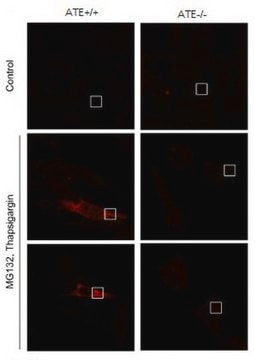
Immunocytochemistry Analysis: 10 µg/mL from a representative lot detected BiP Nt-Glu19 arginylation induction in (18-hr 3 µM MG132 and 200 nM thapsigargin) treated wild-type, but not arginine-tRNA-protein transferase 1/ATE1-deficient, MEFs (Courtesy of Yong Tae Kwon, Ph.D. , Seoul National University, Korea).
Western Blotting Analysis: 0.2 µg/mL from a representative lot detected BiP Nt-Glu19 arginylation induction in (18-hr 3 µM MG132 and 200 nM thapsigargin) treated HeLa cells (Courtesy of Yong Tae Kwon, Ph.D. , Seoul National University, Korea).
Western Blotting Analysis: 0.2 µg/mL from a representative lot detected a target R-BiP(19-651)-GFP fusion band in MEF cells transfected to express Ub-R-BiP(19-651)-GFP or Ub-BiP(19-651)-GFP, but not Ub-V-BiP(19-651)-GFP. In ATE1-deficient MEFs, the target R-BiP(19-651)-GFP band was detected only when the cells were tranfected to express Ub-R-BiP(19-651)-GFP, but not Ub-BiP(19-651)-GFP (Courtesy of Yong Tae Kwon, Ph.D. , Seoul National University, Korea).
Dot Blot Analysis: A representative lot detected the immunogen peptide, but not the control peptide without arginylation at the N-terminal Glu19 (Cha-Molstad, H., et al. (2015). Nat. Cell Biol. 17(7):917-929).
ELISA Analysis: A representative lot detected the immunogen peptide, but not the control peptide without arginylation at the N-terminal Glu19 (Cha-Molstad, H., et al. (2015). Nat. Cell Biol. 17(7):917-929).
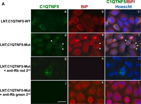
Immunocytochemistry Analysis: A representative lot detected poly(dA:dT) transfection-induced formation of BiP arginylation/R-BiP-positive puncta co-localized with those containing p62, LC3, and ubiquitin conjugates, while R-BiP and ER stainings are mutually exclusive (Cha-Molstad, H., et al. (2015). Nat. Cell Biol. 17(7):917-929).
Western Blotting Analysis: A representative lot detected the production of R-BiP(19-651)-Tag fusions from exogenously expressed Ub-BiP(19-N)-Tag and Ub-R-BiP(19-N)-Tag, but not Ub-V-BiP(20-N)-Tag, constructs. In ATE1-deficient cells, the target R-BiP(19-651)-GFP band was detected only when the cells were tranfected to express Ub-R-BiP(19-651)-GFP, but not Ub-BiP(19-651)-GF (Cha-Molstad, H., et al. (2015). Nat. Cell Biol. 17(7):917-929).
Western Blotting Analysis: A representative lot detected BiP (GRP78) Nt-Glu19 arginylation induction upon arginine-tRNA-protein transferase 1 (ATE1) 1A7A isoform overexpression or transfection of various dsDNAs, including poly(dA:dT), in HeLa cells. Combined proteasome inhibition and ER stress induction by an 18-hr 10 µM MG132 and 100 nM thapsigargin treatment synergized the two drugs′ efficacy toward cellular Calreticulin Nt-Glu18 arginylation induction (Cha-Molstad, H., et al. (2015). Nat. Cell Biol. 17(7):917-929).
Biochem/physiol Actions
Physical form
Preparation Note
Analysis Note
Western Blotting Analysis: 1 µg/mL of this antibody detected BiP (GRP78) Nt-Glu19 arginylation induction in 7.5 µg of lysate from (17-hr 3 µM MG132 and 200 nM thapsigargin) treated HEK293 cells.
Other Notes
Disclaimer
1 of 1
Este artículo | |||
|---|---|---|---|
| Quality Level 100 | Quality Level 200 | Quality Level 100 | Quality Level 100 |
| antibody form affinity isolated antibody | antibody form IgG fraction of antiserum | antibody form affinity isolated antibody | antibody form purified immunoglobulin |
| conjugate unconjugated | conjugate unconjugated | conjugate unconjugated | conjugate unconjugated |
| biological source rabbit | biological source rabbit | biological source rabbit | biological source mouse |
| shipped in ambient | shipped in dry ice | shipped in wet ice | shipped in wet ice |
| UniProt accession no. | UniProt accession no. | UniProt accession no. | UniProt accession no. |

Still not finding the right product?
Pruebe nuestra Herramienta de selección de productos para limitar sus opciones
Clase de almacenamiento
12 - Non Combustible Liquids
wgk
WGK 2
flash_point_f
Not applicable
flash_point_c
Not applicable
Certificados de análisis (COA)
Busque Certificados de análisis (COA) introduciendo el número de lote del producto. Los números de lote se encuentran en la etiqueta del producto después de las palabras «Lot» o «Batch»
¿Ya tiene este producto?
Encuentre la documentación para los productos que ha comprado recientemente en la Biblioteca de documentos.
Número de artículo de comercio global
| SKU | GTIN |
|---|---|
| ABS2103 | 04054839083709 |