Accéder au contenu
Merck

Passer à

MABC1122

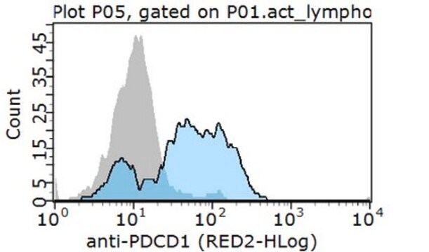
Anti-PD-1 Antibody, clone EH33

clone EH33.1F9.G4.5, from mouse

Synonyme(s) :

Programmed cell death protein 1, hPD-1, CD279

Se connecter pour consulter les tarifs organisationnels et contractuels.

Sélectionner une taille de conditionnement

Changer de vue
ConditionnementRéférenceDisponibilitéPrix
25 μg
Vérifier la disponibilité des articles du panier
159,00 €
100 μg
Vérifier la disponibilité des articles du panier
402,00 €

A propos de cet article

UNSPSC Code:
12352203
NACRES:
NA.41
eCl@ss:
32160702
Conjugate:
unconjugated
Clone:
EH33.1F9.G4.5, monoclonal
Application:
IHC
Citations:
-

159,00 €


Vérifier la disponibilité des articles du panier
Un anticorps recombinant, sans conservateur, est disponible pour votre cible. Essayez ZRB1025

Demander une commande groupée
Service technique
Besoin d'aide ? Notre équipe de scientifiques expérimentés est là pour vous.
Laissez-nous vous aider


biological source

mouse

Quality Segment

conjugate

unconjugated

antibody form

purified immunoglobulin

antibody product type

primary antibodies

clone

EH33.1F9.G4.5, monoclonal

species reactivity

human

packaging

antibody small pack of 25 μg

technique(s)

immunohistochemistry: suitable (paraffin)

isotype

IgG2aκ

NCBI accession no.

UniProt accession no.

target post-translational modification

unmodified

Gene Information

human ... PDCD1(5133)

General description

31.65 kDa calculated.
Programmed cell death protein 1 (UniProt: Q15116; also known as Protein PD-1, hPD-1, CD279) is encoded by the PDCD1 (also known as PD1) gene (Gene ID: 5133) in human. PD-1 is a monomeric inhibitory cell surface receptor involved in the regulation of T-cell function during immunity and tolerance. PD-1 is synthesized with a signal peptide (aa 1-20), which is subsequently cleaved off. The mature form contains an extracellular domain (aa 24-170), a transmembrane domain (aa 171-191), and a cytoplasmic domain (aa 192-288). PD-1 also contains a single N-terminal immunoglobulin variable region (IgV) like domain. PD-1 and its ligands, PD-L1 and PD-L2 play a key role in the maintenance of peripheral tolerance, a process by which the quiescence of autoreactive mature T cells is maintained. However, tumors and pathogens that cause chronic infections can exploit this pathway to escape T-cell mediated tumor-specific and pathogen-specific immunity. The effector functions of T-cells expressing PD-1 can be downregulated by PD-L1 or PD-L2 expressed by the tumor cells. PD-1 lacks SH2- or SH3-binding motifs on its cytoplasmic tail, but contains the N-terminal sequence VDYGEL that forms an immunoreceptor tyrosine-based inhibition motif (V/I/LxYxxL), which recruits SH2 domain-containing phosphatases. The cytoplasmic tail also contains the C-terminal sequence TEYATI, which forms an immunoreceptor tyrosine-based switch motif (TxYxxL). PD-1 ligation is reported to inhibit the activation of T-cell receptor proximal kinases, which results in attenuation of Lck-mediated phosphorylation of ZAP-70 and initiation of downstream events. PD-1 is also reported to impair the activation of the MEK-ERK MAP kinase pathway by inhibiting activation of PLC- 1 and Ras. (Ref.: Boussiotis, VA (2016). N. Engl. J. Med. 375; 1767-1778).

Immunogen

Epitope: extracellular domain
Recombinant protein fragment corresponding to the extracellular domain of human PD-1 (as an Ig fusion protein).

Application

Anti-PD-1, clone EH33, Cat. No. MABC1122, is a mouse monoclonal antibody that detects Programmed cell death protein 1 (PD-1) in human cells and has been tested for use in Immunohistochemistry (Paraffin).
Immunohistochemistry Analysis: A representative lot detected PD-1 in Immunohistochemistry applications (Sridharan, V., et. al. (2016). Cancer Immunol Res. 4(8):679-87; Calles, A., et. al. (2015). J Thorac Oncol. 10(12):1726-35; Bachireddy, P., et. al. (2014). Blood. 123(9):1412-21).
Research Category
Inflammation & Immunology

Biochem/physiol Actions

Clone EH33 specifically detects PD-1 in human cells. It targets an epitope with the extracellular domain.

Physical form

Format: Purified
Protein G purified
Purified mouse monoclonal antibody IgG2a in buffer containing 0.1 M Tris-Glycine (pH 7.4), 150 mM NaCl with 0.05% sodium azide.

Preparation Note

Stable for 1 year at 2-8°C from date of receipt.

Analysis Note

Evaluated by Immunohistochemistry in human tonsil and human bone marrow tissues.

Immunohistochemistry Analysis: A 1:250 dilution of this antibody detected PD-1 in human tonsil and human bone marrow tissues.

Other Notes

Concentration: Please refer to lot specific datasheet.

Disclaimer

Unless otherwise stated in our catalog or other company documentation accompanying the product(s), our products are intended for research use only and are not to be used for any other purpose, which includes but is not limited to, unauthorized commercial uses, in vitro diagnostic uses, ex vivo or in vivo therapeutic uses or any type of consumption or application to humans or animals.

Comparer avec des articles similaires

Voir la comparaison complète

Montrer les différences

1 of 1

Cet article
MABF261SAB3500123MABC1115
biological source

mouse

biological source

mouse

biological source

mouse

biological source

mouse

conjugate

unconjugated

conjugate

unconjugated

conjugate

unconjugated

conjugate

unconjugated

species reactivity

human

species reactivity

human

species reactivity

rat, human, mouse

species reactivity

human

antibody form

purified immunoglobulin

antibody form

purified immunoglobulin

antibody form

purified immunoglobulin

antibody form

purified immunoglobulin

clone

EH33.1F9.G4.5, monoclonal

clone

16A2.1, monoclonal

clone

12A7D7, monoclonal

clone

5H1, monoclonal

Gene Information

human ... PDCD1(5133)

Gene Information

human ... PDCD1(5133)

Gene Information

human ... PDCD1(5133)

Gene Information

human ... CD274(29126)


Still not finding the right product?

Essayez notre Outil de sélection de produits pour affiner vos choix.


Classe de stockage

12 - Non Combustible Liquids

wgk

WGK 1

flash_point_f

Not applicable

flash_point_c

Not applicable


Certificats d'analyse (COA)

Recherchez un Certificats d'analyse (COA) en saisissant le numéro de lot du produit. Les numéros de lot figurent sur l'étiquette du produit après les mots "Lot" ou "Batch".

Déjà en possession de ce produit ?

Retrouvez la documentation relative aux produits que vous avez récemment achetés dans la Bibliothèque de documents.

Consulter la Bibliothèque de documents


Numéro d'article de commerce international

RéférenceGTIN
MABC1122-25UG04054839448324

Questions

Évaluations

Aucune valeur de notation

Filtres actifs