Ugrás a tartalomra
Merck
Összes fotó(1)

Fontos dokumentumok

MABT26

Sigma-Aldrich

Anti-E-cadherin Antibody, clone DECMA-1

clone Decma-1, from rat

Szinonimák:

Anti-Anti-Arc-1, Anti-Anti-BCDS1, Anti-Anti-CD324, Anti-Anti-CDHE, Anti-Anti-ECAD, Anti-Anti-LCAM, Anti-Anti-UVO

Bejelentkezésa Szervezeti és Szerződéses árazás megtekintéséhez


About This Item

UNSPSC kód:
12352203
eCl@ss:
32160702
NACRES:
NA.41
klón:
Decma-1, monoclonal
application:
FACS
ICC
IHC
IP
WB
faj reaktivitás:
human, mouse
technika/technikák:
flow cytometry: suitable
immunocytochemistry: suitable
immunohistochemistry: suitable
immunoprecipitation (IP): suitable
western blot: suitable
citations:
24

biológiai forrás

rat

Minőségi szint

antitest forma

purified antibody

antitest terméktípus

primary antibodies

klón

Decma-1, monoclonal

faj reaktivitás

human, mouse

technika/technikák

flow cytometry: suitable
immunocytochemistry: suitable
immunohistochemistry: suitable
immunoprecipitation (IP): suitable
western blot: suitable

izotípus

IgG1κ

NCBI elérési szám

UniProt elérési szám

kiszállítva

dry ice

célzott transzláció utáni módosítás

unmodified

Géninformáció

human ... CDH1(999)
mouse ... Cdh1(12550)

Related Categories

Általános leírás

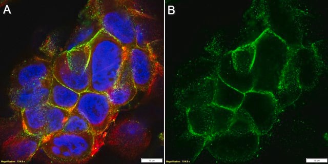
Cadherins are a family of transmembrane glycoproteins involved in Ca2+-dependent cell to cell interactions. The members of the family are differentially expressed in various tissues and function in the maintenance of tissue integrity, morphogenesis, and migration. Cadherins are divided into type I and type II subgroups. Type I cadherins include epithelial cadherin (E-cadherin), neural cadherin (N-cadherin), placental cadherin (P-cadherin) and retinal cadherin (R-cadherin). Type II include kidney cadherin (K-cadherin) and osteoblast cadherin (OB-cadherin). One of the best characterized cadherins is E-cadherin (epithelial cadherin or cadherin-1). It consists of calcium-binding extracellular domains responsible for interactions with other E-cadherin molecules on neighboring cells and interaction with integrin alphaEbeta7. The cytoplasmic domain of E-cadherin is linked to the actin cytoskeleton through cytoplasmic catenin proteins, thus establishing a complex localized to adherens junctions. In carcinomas, E-cadherin is frequently downregulated, consistent with its function of an invasion suppressor in normal epithelia.

Immunogén

Mouse embryonal carcinoma cell line PCC4

Alkalmazás

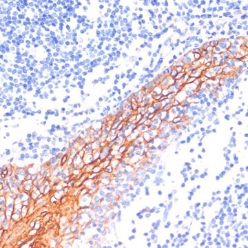
Immunohistochemistry Analysis: 1:300 dilution of this antibody has been shown to detect E-cadherin in mouse GI tract tissue.

Inhibition: A previous lot was used by an independent laboratory in inhibion studies, blocking E-cadherin associated cell adhesion. (Vestweber, 1985)

Flow Cytometry Analysis: A previous lot was used by an independent laboratory in flow cytometry applications. (Tang, 1994)
Research Category
Cell Structure
Research Sub Category
Adhesion (CAMs)
This Anti-E-cadherin Antibody, clone DECMA-1 is validated for use in WB, IH, IC, IP, FC, FUNC for the detection of E-cadherin.

Minőség

Evaluated by Western Blot in A431 cell lysate.

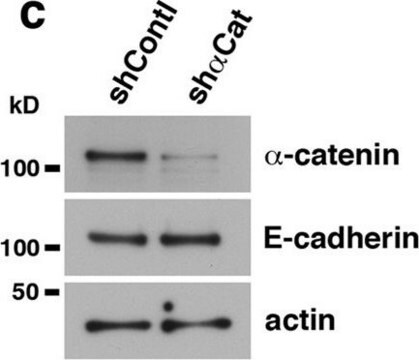
Western Blot Analysis: 1 µg/mL of this antibody detected E-Cadherin in 10 µg of A431 cell lysate.

Cél megnevezése

~ 106 kDa observed MW.

Fizikai forma

Format: Purified
Protein G
Purified rat monoclonal IgG1κ in buffer containing PBS without preservative.

Tárolás és stabilitás

Stable for 1 year at -20°C from date of receipt.
Handling Recommendations: Upon receipt and prior to removing the cap, centrifuge the vial and gently mix the solution. Aliquot into microcentrifuge tubes and store at -20°C. Avoid repeated freeze/thaw cycles, which may damage IgG and affect product performance.

Analízis megjegyzés

Control
A431 cell lysate

Egyéb megjegyzések

Concentration: Please refer to the Certificate of Analysis for the lot-specific concentration.

Jogi nyilatkozat

Unless otherwise stated in our catalog or other company documentation accompanying the product(s), our products are intended for research use only and are not to be used for any other purpose, which includes but is not limited to, unauthorized commercial uses, in vitro diagnostic uses, ex vivo or in vivo therapeutic uses or any type of consumption or application to humans or animals.

Nem találja a megfelelő terméket?  

Próbálja ki a Termékválasztó eszköz. eszközt

Tárolási osztály kódja

12 - Non Combustible Liquids

WGK

WGK 2

Lobbanási pont (F)

Not applicable

Lobbanási pont (C)

Not applicable


Analitikai tanúsítványok (COA)

Analitikai tanúsítványok (COA) keresése a termék sarzs-/tételszámának megadásával. A sarzs- és tételszámok a termék címkéjén találhatók, a „Lot” vagy „Batch” szavak után.

Már rendelkezik ezzel a termékkel?

Az Ön által nemrégiben megvásárolt termékekre vonatkozó dokumentumokat a Dokumentumtárban találja.

Dokumentumtár megtekintése

X Y Zhang et al.
Clinical and experimental immunology, 129(2), 288-296 (2002-08-08)
High levels of interleukin-6 (IL-6) and interleukin-8 (IL-8) have been demonstrated in the peritoneal fluid of benign and malignant gynaecological disease. Peritoneal monocytes and macrophages, endometrial cells, endometrial and peritoneal stromal cells and tumour cells produce these cytokines in vitro.
Jacopo Di Russo et al.
eLife, 10 (2021-09-24)
Nanometer-scale properties of the extracellular matrix influence many biological processes, including cell motility. While much information is available for single-cell migration, to date, no knowledge exists on how the nanoscale presentation of extracellular matrix receptors influences collective cell migration. In
Marianela G Dalghi et al.
JCI insight, 6(19) (2021-09-01)
The mechanisms that link visceral mechanosensation to the perception of internal organ status (i.e., interoception) remain elusive. In response to bladder filling, the urothelium releases ATP, which is hypothesized to stimulate voiding function by communicating the degree of bladder fullness
Nicholas F Dybdal-Hargreaves et al.
Oncotarget, 10(45), 4719-4720 (2019-08-07)
[This corrects the article DOI: 10.18632/oncotarget.23798.].
Pei-Pei Cao et al.
Oncology reports, 43(2), 662-670 (2020-01-03)
Cullin 4A (CUL4A) is a member of the cullin family of proteins and has been demonstrated to be abnormally expressed in various types of malignancies. However, the function of CUL4A in metastasis of lung adenocarcinoma to the bone has rarely

Tudóscsoportunk valamennyi kutatási területen rendelkezik tapasztalattal, beleértve az élettudományt, az anyagtudományt, a kémiai szintézist, a kromatográfiát, az analitikát és még sok más területet.

Lépjen kapcsolatba a szaktanácsadással