Skip to Content
Merck
All Photos(1)

Documents

MABS409

Sigma-Aldrich

Anti-NFATc1 Antibody, clone 7A6

clone 7A6, from mouse

Synonym(s):

Nuclear factor of activated T-cells, cytoplasmic 1, NF-ATc1, NFATc1, NFAT transcription complex cytosolic component, NF-Atc, NFATc

Sign Into View Organizational & Contract Pricing


About This Item

UNSPSC Code:
12352203
eCl@ss:
32160702
NACRES:
NA.41

biological source

mouse

antibody form

purified immunoglobulin

antibody product type

primary antibodies

clone

7A6, monoclonal

species reactivity

human, mouse

technique(s)

flow cytometry: suitable
immunofluorescence: suitable
western blot: suitable

isotype

IgG1

UniProt accession no.

shipped in

wet ice

target post-translational modification

unmodified

Gene Information

human ... NFATC1(4772)

General description

NFATc1, also known as Nuclear factor of activated T-cells, cytoplasmic, calcineurin-dependent 1, is a component of the nuclear factor of activated T cells DNA-binding transcription complex which consists of at least two components: a preexisting cytosolic component that translocates to the nucleus upon T cell receptor (TCR) stimulation, and an inducible nuclear component. This protein plays a main role in the inducible expression of cytokine genes in T-cells, especially in the induction of the IL-2 or IL-4 gene transcription and functions as a major molecular target for the immunosuppressive drugs such as cyclosporine A. NFATc1 is expressed in most human primary lymphocytes and mature human T- and B-cell neoplasms.

Immunogen

Recombinant protein of human NFATc1 amino acids 197-304

Application

Detect NFAT using this mouse monoclonal antibody, Anti-NFATc1 Antibody, clone 7A6 validated for use in western blotting, Flow Cytometry & Immunofluorescence.
Flow Cytometry Analysis: A representative lot detected human NFATc1 in Flow Cytometry. (Brandt C, et al. 2010.)

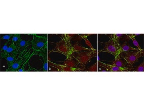
Immunofluorescent Analysis: A representative lot detected human NFATc1 in Immunofluorescence. (Timmerman LA, et al. 1997.)

Quality

Evaluated by Western Blotting in Jurkat, human Th1, and mouse Th1 cell extracts.

Western Blotting Analysis: 1 μg/mL of this antibody detected in Jurkat, human Th1, and mouse Th1 cell extracts.

Target description

~~95, ~110 and ~130 kDa observed

Physical form

Format: Purified
Purified mouse monoclonal IgG1 antibody in buffer containing PBS (pH 7.2) with up to 0.1% sodium azide.

Storage and Stability

The antibody solution should be stored undiluted at 2-8°C and protected from prolonged exposure to light. Do not freeze.

Other Notes

Concentration: Please refer to the Certificate of Analysis for the lot-specific concentration.

Not finding the right product?  

Try our Product Selector Tool.

Storage Class Code

12 - Non Combustible Liquids

WGK

WGK 2

Flash Point(F)

Not applicable

Flash Point(C)

Not applicable


Certificates of Analysis (COA)

Search for Certificates of Analysis (COA) by entering the products Lot/Batch Number. Lot and Batch Numbers can be found on a product’s label following the words ‘Lot’ or ‘Batch’.

Already Own This Product?

Find documentation for the products that you have recently purchased in the Document Library.

Visit the Document Library

Krithika Ramachandran et al.
PLoS biology, 17(10), e3000467-e3000467 (2019-10-08)
Skeletal muscles consist of fibers of differing metabolic activities and contractility, which become remodeled in response to chronic exercise, but the epigenomic basis for muscle identity and adaptation remains poorly understood. Here, we used chromatin immunoprecipitation sequencing of dimethylated histone
Celestia Fang et al.
Journal of immunology (Baltimore, Md. : 1950), 196(11), 4452-4456 (2016-05-18)
Osteoclasts are resorptive cells that are important for homeostatic bone remodeling and pathological bone resorption. Emerging evidence suggests an important role for epigenetic mechanisms in osteoclastogenesis. A recent study showed that epigenetic silencing of the negative regulator of osteoclastogenesis Irf8
Novel Bruton's tyrosine kinase inhibitor TAS5315 suppresses the progression of inflammation and joint destruction in rodent collagen-induced arthritis.
Akasaka, et al.
PLoS ONE, 18, e0282117-e0282117 (2023)
Shi-Ying Cai et al.
Journal of hepatology, 74(3), 550-559 (2020-10-12)
The nuclear factor of activated T-cells (NFAT) plays an important role in immune responses by regulating the expression of inflammatory genes. However, it is not known whether NFAT plays any role in the bile acid (BA)-induced hepatic inflammatory response. Thus
Amado Carreras-Sureda et al.
eLife, 10 (2021-12-17)
Efficient immune responses require Ca2+ fluxes across ORAI1 channels during engagement of T cell receptors (TCR) at the immune synapse (IS) between T cells and antigen presenting cells. Here, we show that ZDHHC20-mediated S-acylation of the ORAI1 channel at residue

Our team of scientists has experience in all areas of research including Life Science, Material Science, Chemical Synthesis, Chromatography, Analytical and many others.

Contact Technical Service