
F4512
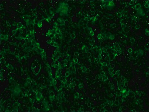
Anti-Human IgG (whole molecule)−FITC antibody produced in rabbit
IgG fraction of antiserum, buffered aqueous solution
Synonym(s):
Rabbit Anti-Human IgG (whole molecule)−Fluorescein isothiocyanate
Select a Size
| Pack Size | SKU | Availability | Price |
|---|
About This Item
biological source
rabbit
Quality Segment
conjugate
FITC conjugate
antibody form
IgG fraction of antiserum
antibody product type
secondary antibodies
clone
polyclonal
form
buffered aqueous solution
storage condition
protect from light
technique(s)
immunofluorescence: 1:64-1:128 using Hep2 cells
storage temp.
−20°C
target post-translational modification
unmodified
General description
Immunogen
Application
Immunofluorescence (1 paper)
Physical form
Disclaimer
1 of 1
This Item | |||
|---|---|---|---|
| biological source rabbit | biological source rabbit | biological source rabbit | biological source goat |
| antibody form IgG fraction of antiserum | antibody form IgG fraction of antiserum | antibody form IgG fraction of antiserum | antibody form IgG fraction of antiserum |
| conjugate FITC conjugate | conjugate FITC conjugate | conjugate FITC conjugate | conjugate FITC conjugate |
| technique(s) immunofluorescence: 1:64-1:128 using Hep2 cells | technique(s) ANA-indirect immunofluorescence: 1:128, direct immunofluorescence: 1:80 | technique(s) immunohistochemistry (formalin-fixed, paraffin-embedded sections): 1:100, indirect immunofluorescence: 1:100 | technique(s) immunofluorescence: 1:32-1:64 using Hep2 cells |
| form buffered aqueous solution | form buffered aqueous solution | form buffered aqueous solution | form buffered aqueous solution |
| storage temp. −20°C | storage temp. −20°C | storage temp. −20°C | storage temp. −20°C |


Still not finding the right product?
Try our Product Selector Tool to narrow your options
Storage Class
10 - Combustible liquids
wgk
nwg
flash_point_f
Not applicable
flash_point_c
Not applicable
ppe
Eyeshields, Gloves, multi-purpose combination respirator cartridge (US)
Choose from one of the most recent versions:
Already Own This Product?
Find documentation for the products that you have recently purchased in the Document Library.
Related Content
Global Trade Item Number
| SKU | GTIN |
|---|---|
| F4512-1ML | 04061833617762 |
| F4512-2ML | 04061833617779 |