
05-806
Anti-phospho-Histone H3 (Ser10) Antibody, clone 3H10
clone 3H10, Upstate®, from mouse
Szinonimák:
H3 histone, family 3A, H3S10P, Histone H3 (phospho S10), H3 histone, family 3A, H3 histone, family 3B, H3 histone, family 3B (H3.3B)
About This Item
Javasolt termékek
biológiai forrás
mouse
Minőségi szint
antitest forma
purified immunoglobulin
antitest terméktípus
primary antibodies
klón
3H10, monoclonal
faj reaktivitás
human
gyártó/kereskedő neve
Upstate®
technika/technikák
flow cytometry: suitable
immunocytochemistry: suitable
immunofluorescence: suitable
inhibition assay: suitable (peptide)
multiplexing: suitable
western blot: suitable
izotípus
IgG1κ
NCBI elérési szám
UniProt elérési szám
kiszállítva
wet ice
célzott transzláció utáni módosítás
phosphorylation (pSer10)
Géninformáció
human ... HIST1H3F(8968)
Related Categories
Általános leírás
Egyediség
Alkalmazás
This antibody has been reported by an independent laboratory to detect phosphorylated histone H3 using flow cytometry.
Beadlyte Histone-Peptide Specificity Assay:
1:1,000 to 1:81,000 dilutions of a previous lot were incubated with histone H3 peptides containing various modifications conjugated to Luminex microspheres. (Figure B). Only the peptide containing phospho-serine 10 was detected.
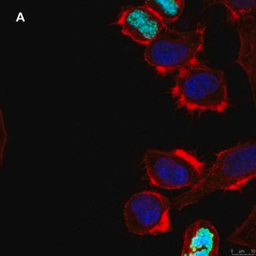
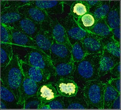
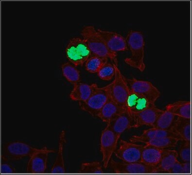
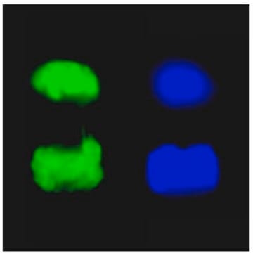
Immunocytochemistry:
0.2 μg/mL of a previous lot showed positive chromosome immunostaining for mitotic A431 and HeLa cells fixed with 95% ethanol and 5% acetic acid and permeabilized with 0.1% Triton X-100.
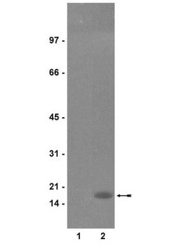
Peptide Inhibition Analysis:
Detection of histone H3 in immunoblots of colcemid treated HeLa acid extracts by anti-phospho-Histone H3 (Ser10) was diminished by 10 μM of histone H3 peptide containing phospho-serine 10, but not by peptides containing phospho-serine 28 or an unmodified histone H3 sequence (Figure C).
Immunofluorescence:
Minőség
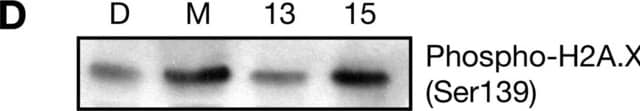
Western Blot Analysis:
0.05-0.5 μg/mL of this lot detected phosphorylated histone H3 in acid extracted proteins from mitotic HeLa cells treated with colcemid (Catalog # 17-306), but not unmodified recombinant Histone H3 (Catalog # 14-494) (Figure A).
Cél megnevezése
Fizikai forma
Tárolás és stabilitás
Handling Recommendations:
Upon receipt, and prior to removing the cap, centrifuge the vial and gently mix the solution. Aliquot into microcentrifuge tubes and store at -20°C. Avoid repeated freeze/thaw cycles, which may damage IgG and affect product performance. Note: Variability in freezer temperatures below -20°C may cause glycerol containing solutions to become frozen during storage.
Analízis megjegyzés
UV-treated 293 cell extracts, UV-treated HeLa cell extracts, breast cancer tissue, HEPG2 cell extracts.
Egyéb megjegyzések
Jogi információk
Nem találja a megfelelő terméket?
Próbálja ki a Termékválasztó eszköz. eszközt
Tárolási osztály kódja
12 - Non Combustible Liquids
WGK
WGK 1
Lobbanási pont (F)
Not applicable
Lobbanási pont (C)
Not applicable
Analitikai tanúsítványok (COA)
Analitikai tanúsítványok (COA) keresése a termék sarzs-/tételszámának megadásával. A sarzs- és tételszámok a termék címkéjén találhatók, a „Lot” vagy „Batch” szavak után.
Már rendelkezik ezzel a termékkel?
Az Ön által nemrégiben megvásárolt termékekre vonatkozó dokumentumokat a Dokumentumtárban találja.
Tudóscsoportunk valamennyi kutatási területen rendelkezik tapasztalattal, beleértve az élettudományt, az anyagtudományt, a kémiai szintézist, a kromatográfiát, az analitikát és még sok más területet.
Lépjen kapcsolatba a szaktanácsadással