Ugrás a tartalomra
Merck

C7974

Sigma-Aldrich

Monoclonal Anti-Collagen, Type X antibody produced in mouse

clone COL-10, ascites fluid

Szinonimák:

Anti-Col10, Anti-Col10a-1

Bejelentkezésa Szervezeti és Szerződéses árazás megtekintéséhez


About This Item

MDL-szám:
UNSPSC kód:
12352203
NACRES:
NA.41

biológiai forrás

mouse

Minőségi szint

konjugátum

unconjugated

antitest forma

ascites fluid

antitest terméktípus

primary antibodies

klón

COL-10, monoclonal

molekulatömeg

antigen 60 kDa (in denatured-reduced preparations)

tartalmaz

15 mM sodium azide

faj reaktivitás

deer, human, pig

technika/technikák

dot blot: suitable
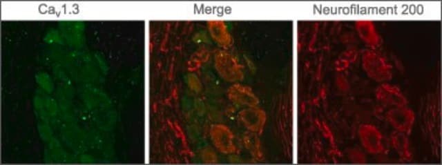
immunocytochemistry: 1:1,000 using HT 1080 human fibrosarcoma cells
western blot: suitable

izotípus

IgM

UniProt elérési szám

kiszállítva

dry ice

tárolási hőmérséklet

−20°C

célzott transzláció utáni módosítás

unmodified

Géninformáció

human ... COL10A1(1300)

Related Categories

Általános leírás

Collagens are extracellular glycoproteins made up of three polypeptides that intermingle to form a triple helix. Type X collagen is a homotrimer of 59 kD α1(X) chains found in fetal hypertrophic cartilage in the growth zones of long bones, vertebrae and ribs, whereas in adults it is also present in thyroid cartilage. Monoclonal anti-collagen, type X antibody can be used in histological and immunohistochemical evaluation of cell cultures. It is also useful in study of specific differential tissue expression.
Monoclonal Anti-Collagen, Type X (mouse IgM isotype) is derived from the COL-10 hybridoma produced by the fusion of mouse myeloma cells and splenocytes from a BALB/c mouse immunized with porcine collagen type X. Collagen type X shares a similar domain structure with type VIII collagen: a central triple-helical (COL1) domain of 50 kDa is flanked by N-terminal (NC2) and C-terminal (NC1) non-triple-helical domains.

Immunogén

purified pig collagen type X.

Alkalmazás

Monoclonal Anti-Collagen, Type X antibody produced in mouse has been used in
  • enzyme linked immunosorbent assay (ELISA)
  • dot-blot
  • immunoblotting and
  • immunohistochemistry

Biokémiai/fiziológiai hatások

Type X collagen is a product of hypertrophic chondrocytes. Type X collagen is non-fibrillar, but forms fine pericellular filaments in association with cartilage collagen. It interacts with matrix proteins, such as connexin V, chondrocalcein, collagen II and proteoglycans, as well as with Ca2+. It acts as a scaffold to avoid local collapse during endochondral ossification and also has a role in cartilage mineralization. Mutations in the NC1-encoding domain of the human α1(X) collagen gene, are associated with Schmid metaphysical chondroplasia.

Jogi nyilatkozat

Unless otherwise stated in our catalog or other company documentation accompanying the product(s), our products are intended for research use only and are not to be used for any other purpose, which includes but is not limited to, unauthorized commercial uses, in vitro diagnostic uses, ex vivo or in vivo therapeutic uses or any type of consumption or application to humans or animals.

Nem találja a megfelelő terméket?  

Próbálja ki a Termékválasztó eszköz. eszközt

Tárolási osztály kódja

10 - Combustible liquids

WGK

WGK 3

Lobbanási pont (F)

Not applicable

Lobbanási pont (C)

Not applicable


Válasszon a legfrissebb verziók közül:

Analitikai tanúsítványok (COA)

Lot/Batch Number

Nem találja a megfelelő verziót?

Ha egy adott verzióra van szüksége, a tétel- vagy cikkszám alapján rákereshet egy adott tanúsítványra.

Már rendelkezik ezzel a termékkel?

Az Ön által nemrégiben megvásárolt termékekre vonatkozó dokumentumokat a Dokumentumtárban találja.

Dokumentumtár megtekintése

Schmid type of metaphyseal chondrodysplasia and COL10A1 mutations?findings in 10 patients
Makitie O, et al.
American Journal of Medical Genetics. Part A, 137(3), 241-248 (2005)
Christopher R Rowland et al.
Biomaterials, 91, 57-72 (2016-03-22)
The native extracellular matrix of cartilage contains entrapped growth factors as well as tissue-specific epitopes for cell-matrix interactions, which make it a potentially attractive biomaterial for cartilage tissue engineering. A limitation to this approach is that the native cartilage extracellular
Temporal and spatial modulation of chondrogenic foci in subchondral microdrill holes by chitosan-glycerol phosphate/blood implants
Chevrier A, et al.
Osteoarthritis and Cartilage, 19(1), 136-144 (2011)
Direct bone morphogenetic protein 2 and Indian hedgehog gene transfer for articular cartilage repair using bone marrow coagulates
Sieker JT, et al.
Osteoarthritis and Cartilage, 23(3), 433-442 (2015)
Aasma A Khan et al.
Biotechnology progress, 25(2), 508-515 (2009-03-19)
The use of bioreactors for cartilage tissue engineering has become increasingly important as traditional batch-fed culture is not optimal for in vitro tissue growth. Most tissue engineering bioreactors rely on convection as the primary means to provide mass transfer; however

Tudóscsoportunk valamennyi kutatási területen rendelkezik tapasztalattal, beleértve az élettudományt, az anyagtudományt, a kémiai szintézist, a kromatográfiát, az analitikát és még sok más területet.

Lépjen kapcsolatba a szaktanácsadással