Ugrás a tartalomra
Merck
Összes fotó(3)

Fontos dokumentumok

MABN495

Sigma-Aldrich

Anti-Aldh1L1 Antibody, clone N103/39

clone N103/39, from mouse

Szinonimák:

Cytosolic 10-formyltetrahydrofolate dehydrogenase, 10-FTHFDH, FDH, Aldehyde dehydrogenase family 1 member L1

FBP-CI

Bejelentkezésa Szervezeti és Szerződéses árazás megtekintéséhez


About This Item

UNSPSC kód:
12352203
eCl@ss:
32160702
NACRES:
NA.41

biológiai forrás

mouse

Minőségi szint

antitest forma

purified immunoglobulin

antitest terméktípus

primary antibodies

klón

N103/39, monoclonal

faj reaktivitás

human, rat, mouse

technika/technikák

immunofluorescence: suitable
immunohistochemistry: suitable
western blot: suitable

izotípus

IgG1κ

NCBI elérési szám

UniProt elérési szám

kiszállítva

wet ice

célzott transzláció utáni módosítás

unmodified

Géninformáció

human ... ALDH1L1(10840)

Általános leírás

Aldh1L1, also known as 10-formyltetrahydrofolate dehydrogenase (10-FTHFDH), is a multidomain protein that is found in liver cytosol and also serves as a CNS astrocyte marker. Aldh1L1 is comprised of two functionally unrelated domains, an aldehyde dehydrogenase-homologous domain and a folate-binding hydrolase domain. These two domains are thought to be connected by a 100-residue linker. It is known to induce phosphorylation of p53 at Ser6, which is a critical step in stimulating apoptosis.

Egyediség

This Aldh1L1 monoclonal antibody does not exhibit cross-reactivity toward Aldh1L2.

Immunogen

Recombinant full-length rat Aldh1L1.

Alkalmazás

Anti-Aldh1L1, clone N103/39 is an antibody targeting the Aldh1L1 protein, validated for use in Immunofluorescence, Immunohistochemistry, and Western Blotting.
Research Category
Neuroscience
Research Sub Category
Sensory & PNS
Western Blot Analysis: 0.5 µg/mL from a representative lot detected Aldh1L1 in 10 µg of human brain tissue lysate.

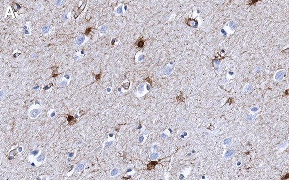
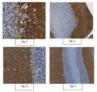
Immunohistochemistry Analysis: A 1:2,000 dilution from a representative lot detected Aldh1L1 rat cerebral cortex tissue and human pons/midbrain tissue.

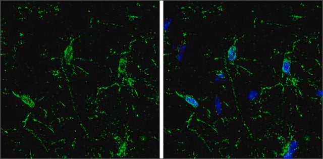
Immunofluorescence Analysis: A representative lot detected Aldh1L1 in rat cortex and cerebellum tissue.

Minőség

Evaluated by Western Blot in mouse brain tissue lysate.

Western Blot Analysis: 0.5 µg/mL of this antibody detected Aldh1L1 in 10 µg of mouse brain tissue lysate.

Cél megnevezése

~99 kDa observed

Fizikai forma

Format: Purified
Protein G Purified
Purified mouse IgG1κ in buffer containing 0.1 M Tris-Glycine (pH 7.4), 150 mM NaCl with 0.05% sodium azide.

Tárolás és stabilitás

Stable for 1 year at 2-8°C from date of receipt.

Analízis megjegyzés

Control
Mouse brain tissue lysate

Egyéb megjegyzések

Concentration: Please refer to the Certificate of Analysis for the lot-specific concentration.

Jogi nyilatkozat

Unless otherwise stated in our catalog or other company documentation accompanying the product(s), our products are intended for research use only and are not to be used for any other purpose, which includes but is not limited to, unauthorized commercial uses, in vitro diagnostic uses, ex vivo or in vivo therapeutic uses or any type of consumption or application to humans or animals.

Nem találja a megfelelő terméket?  

Próbálja ki a Termékválasztó eszköz. eszközt

Tárolási osztály kódja

12 - Non Combustible Liquids

WGK

WGK 1

Lobbanási pont (F)

Not applicable

Lobbanási pont (C)

Not applicable


Analitikai tanúsítványok (COA)

Analitikai tanúsítványok (COA) keresése a termék sarzs-/tételszámának megadásával. A sarzs- és tételszámok a termék címkéjén találhatók, a „Lot” vagy „Batch” szavak után.

Már rendelkezik ezzel a termékkel?

Az Ön által nemrégiben megvásárolt termékekre vonatkozó dokumentumokat a Dokumentumtárban találja.

Dokumentumtár megtekintése

Sheila Espírito-Santo et al.
Glia, 69(6), 1429-1443 (2021-01-27)
Central nervous system (CNS) function depends on precise synaptogenesis, which is shaped by environmental cues and cellular interactions. Astrocytes are outstanding regulators of synapse development and plasticity through contact-dependent signals and through the release of pro- and antisynaptogenic factors. Conversely
New ?- and ?-synuclein immunopathological lesions in human brain.
Surgucheva, I; Newell, KL; Burns, J; Surguchov, A
Acta Neuropathologica Communications null
Jeremy S Rabinowitz et al.
Proceedings of the National Academy of Sciences of the United States of America, 114(5), E717-E726 (2017-01-18)
Regeneration requires cells to regulate proliferation and patterning according to their spatial position. Positional memory is a property that enables regenerating cells to recall spatial information from the uninjured tissue. Positional memory is hypothesized to rely on gradients of molecules
Maria de Los Angeles Castillo-Rodriguez et al.
Histochemistry and cell biology, 158(1), 15-38 (2022-04-06)
Myelin loss with consecutive axon degeneration and impaired remyelination are the underlying causes of progressive disease in patients with multiple sclerosis. Astrocytes are suggested to play a major role in these processes. The unmasking of distinct astrocyte identities in health
Krishna C Vadodaria et al.
Stem cell reports, 16(4), 825-835 (2021-03-06)
Bipolar disorder (BD) is characterized by cyclical mood shifts. Studies indicate that BD patients have a peripheral pro-inflammatory state and alterations in glial populations in the brain. We utilized an in vitro model to study inflammation-related phenotypes of astrocytes derived from

Tudóscsoportunk valamennyi kutatási területen rendelkezik tapasztalattal, beleértve az élettudományt, az anyagtudományt, a kémiai szintézist, a kromatográfiát, az analitikát és még sok más területet.

Lépjen kapcsolatba a szaktanácsadással