MAB3420
ANTI-TAU (MAPT) Antibody
CHEMICON®, mouse monoclonal, PC1C6
Szinonimák:
Anti-Tau Antibody
About This Item
Javasolt termékek
Terméknév
Anti-Tau-1 Antibody, clone PC1C6, clone PC1C6, Chemicon®, from mouse
biológiai forrás
mouse
Minőségi szint
antitest forma
purified antibody
klón
PC1C6, monoclonal
faj reaktivitás
human, rat, bovine
kiszerelés
antibody small pack of 25 μg
gyártó/kereskedő neve
Chemicon®
technika/technikák
immunofluorescence: suitable
immunohistochemistry: suitable
western blot: suitable
izotípus
IgG2a
NCBI elérési szám
UniProt elérési szám
kiszállítva
dry ice
tárolási hőmérséklet
−20°C
célzott transzláció utáni módosítás
unmodified
Related Categories
Általános leírás
Egyediség
Immunogén
Alkalmazás
Neuroscience
Neurodegenerative Diseases
Immunofluorescence: A 1:1000 dilution of this antibody detected Tau in mouse primary neurons. (Basnet, N., et al. (2018). Nat. Cell Biol. 20(10); 1172-1180.
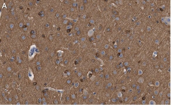
Immunohistochemistry: 5 μg/mL; stains axons in tissue primarily, however in culture Tau expression is not restricted to just axons.
Optimal working dilutions must be determined by end user.
Immunohistochemistry Protocol
Dephosphorylation of tissue sections (optional)
Dephosphorylation with alkaline phosphatase is recommended for staining neurofibrillary tangles in Alzheimer′s brain tissue with anti-tau-1 (6). This treatment changes the staining pattern of anti-tau-1 to include cell bodies, dendrites and axons of neurons. In untreated samples, anti-tau-1 stains axons only.
1. Incubate tissue sections at +32°C for 2.5 hours with constant agitation in the following solution: 100 mM Tris-HCl, pH 8.0; 130 units/mL alkaline phosphatase, 1 mM PMSF, 10 μg/mL pepstatin and 10 μg/mL leupeptin.
2. Rinse sections twice, 3 min per rinse, with 100 mM Tris-HCl, pH 8.0.
Anti-tau-1 staining
1. Block non-specific binding by incubating sections in PBS containing 1% (v/v) normal animal serum, and 0.03% (w/v) Triton X-100. The animal serum should be from the same species as the secondary antibody.
2. Rinse 3 times with PBS, 3 min per rinse.
3. Incubate sections with anti-tau-1, approximately 5 μg/mL, diluted in PBS containing 1% (v/v) normal animal serum.
4. Wash with PBS, changing the solution 3 times over a 3 min period.
5. Detect with a standard secondary antibody detection system (10-13).
Cél megnevezése
Kapcsolódás
Fizikai forma
Tárolás és stabilitás
Analízis megjegyzés
Alzheimer′s brain tissue (dephosphorylation with alkaline phosphatase is recommended for staining neurofibrillary tangles in Alzheimer’s brain tissue) or human T98G glioblastoma cells
Egyéb megjegyzések
Jogi információk
Jogi nyilatkozat
javasolt
Tárolási osztály kódja
12 - Non Combustible Liquids
WGK
WGK 2
Lobbanási pont (F)
Not applicable
Lobbanási pont (C)
Not applicable
Analitikai tanúsítványok (COA)
Analitikai tanúsítványok (COA) keresése a termék sarzs-/tételszámának megadásával. A sarzs- és tételszámok a termék címkéjén találhatók, a „Lot” vagy „Batch” szavak után.
Már rendelkezik ezzel a termékkel?
Az Ön által nemrégiben megvásárolt termékekre vonatkozó dokumentumokat a Dokumentumtárban találja.
Az ügyfelek ezeket is megtekintették
Cikkek
Az immunfluoreszcencia antitesthez kötött fluoreszcens molekulákat használ a fehérjék lokalizációjára, a módosítások megerősítésére és a fehérjekomplexek vizualizálására.
Immunofluorescence uses antibody-conjugated fluorescent molecules for protein localization, modification confirmation, and protein complex visualization.
Derivation and characterization of functional human neural stem cell derived oligodendrocyte progenitor cells (OPCs) that efficiently myelinate primary neurons in culture.
Protocols
Tips and troubleshooting for FFPE and frozen tissue immunohistochemistry (IHC) protocols using both brightfield analysis of chromogenic detection and fluorescent microscopy.
Tudóscsoportunk valamennyi kutatási területen rendelkezik tapasztalattal, beleértve az élettudományt, az anyagtudományt, a kémiai szintézist, a kromatográfiát, az analitikát és még sok más területet.
Lépjen kapcsolatba a szaktanácsadással


