Ugrás a tartalomra
Merck
Összes fotó(3)

Fontos dokumentumok

ABN2192

Sigma-Aldrich

Anti-Calbindin D-28K

from rabbit

Szinonimák:

Calbindin, Calbindin D28, D-28K, PCD-29, Spot 35 protein, Vitamin D-dependent calcium-binding protein

Bejelentkezésa Szervezeti és Szerződéses árazás megtekintéséhez


About This Item

UNSPSC kód:
12352203
eCl@ss:
32160702
NACRES:
NA.41
klón:
polyclonal
application:
ICC
IHC
WB
faj reaktivitás:
mouse, human, rat
technika/technikák:
immunocytochemistry: suitable
immunohistochemistry: suitable (paraffin)
western blot: suitable
citations:
5

biológiai forrás

rabbit

Minőségi szint

antitest forma

affinity isolated antibody

antitest terméktípus

primary antibodies

klón

polyclonal

faj reaktivitás

mouse, human, rat

kiszerelés

antibody small pack of 25 μL

technika/technikák

immunocytochemistry: suitable
immunohistochemistry: suitable (paraffin)
western blot: suitable

NCBI elérési szám

UniProt elérési szám

célzott transzláció utáni módosítás

unmodified

Géninformáció

human ... CALB1(793)
mouse ... Calb1(12307)

Általános leírás

Calbindin (UniProt: P12658; also known as Calbindin D28, D-28K, PCD-29, Spot 35 protein, Vitamin D-dependent calcium-binding protein, avian-type) is encoded by the calb1 gene (gene ID: 12307) in murine species. Calbindin D-28k is a member of a large family of intracellular calcium-binding proteins of the EF-hand related to calmodulin and troponin-C. Calbindin D-28K has four functional calcium-binding sites (aa 24-35; 111-122; 155-166; and 199-210). It has moderate cytoplasmic mobility and comprises six EF-hands, of which one or two are non-functioning in metal binding. EF-2 is viewed as non-metal binding and EF-6 may have a very low Ca2+ affinity. Four EF hands function in calcium binding with rapid to intermediate kinetics and affinity. The biological roles of Calbindin D-28K include calcium regulation and signaling, including calcium transport and uptake, calcification of bone and teeth, and calcium related signaling in neurons and transiently in embryological development. It also protects neurons from apoptotic cell death. Decline in Calbindin D-28K levels in hippocampal dentate granule cells correlates with the kindling model for epilepsy. Calbindin D-28K immunoreactivity is shown to be specifically absent from neuronal populations lost early in ALS, while substantial loss of neurons, which do contain this protein occur in patients with Huntington′s disease. (Ref.: Schmidt, H (2012). Front. Mol. Neurosci. 5; 25).

Egyediség

This rabbit polyclonal antibody detects Calbindin D-28K in Human, Murine, and Rat cells. It targets an epitope within 107 amino acids from the C-terminal region.

Immunogén

Epitope: C-terminus
GST/His-tagged recombinant fragment corresponding to 107 amino acids from the C-terminal end of murine Calbindin D-28K.

Alkalmazás

Anti-Calbindin D-28K Antibody, Cat. No. ABN2192, is a highly specific rabbit polyclonal antibody that targets Calbindin D and has been tested for use in Immunocytochemistry, Immunohistochemistry (Paraffin), and Western Blotting.
Immunocytochemistry Analysis: A 1:100 dilution from a representative lot detected Calbindin D-28K in E18 rat cortical cells.

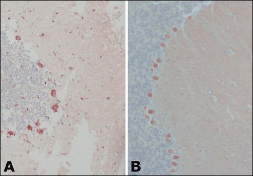
Immunohistochemistry Analysis: A 1:1,000 dilution from a representative lot detected Calbindin D-28K in mouse kidney and human cerebellum tissue sections.
Research Category
Neuroscience

Minőség

Evaluated by Western Blotting in human brain tissue lysate.

Western Blotting Analysis: A 1:500 dilution of this antibody detected Calbindin D-28K in human brain tissue lysate.

Cél megnevezése

~28 kDa observed; 29.99 kDa calculated. Uncharacterized bands may be observed in some lysate(s).

Fizikai forma

Affinity Purified
Format: Purified
Purified rabbit polyclonal antibody in buffer containing 0.1 M Tris-Glycine (pH 7.4), 150 mM NaCl with 0.05% sodium azide.

Tárolás és stabilitás

Stable for 1 year at 2-8°C from date of receipt.

Egyéb megjegyzések

Concentration: Please refer to lot specific datasheet.

Jogi nyilatkozat

Unless otherwise stated in our catalog or other company documentation accompanying the product(s), our products are intended for research use only and are not to be used for any other purpose, which includes but is not limited to, unauthorized commercial uses, in vitro diagnostic uses, ex vivo or in vivo therapeutic uses or any type of consumption or application to humans or animals.

Nem találja a megfelelő terméket?  

Próbálja ki a Termékválasztó eszköz. eszközt

Tárolási osztály kódja

12 - Non Combustible Liquids

WGK

WGK 1

Lobbanási pont (F)

Not applicable

Lobbanási pont (C)

Not applicable


Analitikai tanúsítványok (COA)

Analitikai tanúsítványok (COA) keresése a termék sarzs-/tételszámának megadásával. A sarzs- és tételszámok a termék címkéjén találhatók, a „Lot” vagy „Batch” szavak után.

Már rendelkezik ezzel a termékkel?

Az Ön által nemrégiben megvásárolt termékekre vonatkozó dokumentumokat a Dokumentumtárban találja.

Dokumentumtár megtekintése

Yuxuan Meng et al.
Methods in molecular biology (Clifton, N.J.), 2800, 67-74 (2024-05-06)
The study of cell signaling within tissues can be enhanced using highly multiplexed immunohistochemistry to localize the presence and spatial distribution of numerous pathways of interest simultaneously. Additional data can also be gained by placing the identified proteins into the
Hyun Seung Shin et al.
International journal of molecular sciences, 24(5) (2023-03-12)
Early life stress (ELS) in developing children has been linked to physical and psychological sequelae in adulthood. In the present study, we investigated the effects of ELS on brain and behavioral development by establishing a novel ELS model that combined
Sahithi Attaluri et al.
Frontiers in aging neuroscience, 15, 1200445-1200445 (2023-07-10)
Extracellular vesicles (EVs) released by human-induced pluripotent stem cell (hiPSC)-derived neural stem cells (NSCs) have robust antiinflammatory and neurogenic properties due to therapeutic miRNAs and proteins in their cargo. Hence, hiPSC-NSC-EVs are potentially an excellent biologic for treating neurodegenerative disorders
Song Li et al.
Cell reports, 41(13), 111880-111880 (2022-12-29)
Aging causes an irreversible, cumulative decline in neuronal function. Using the visual system as a model, we show that astrocytes play a critical role in maintaining retinal ganglion cell health and that deletion of SPP1 (secreted phosphoprotein 1, or osteopontin)
Abeer Dagra et al.
NPJ Parkinson's disease, 7(1), 76-76 (2021-08-20)
Pathophysiological damages and loss of function of dopamine neurons precede their demise and contribute to the early phases of Parkinson's disease. The presence of aberrant intracellular pathological inclusions of the protein α-synuclein within ventral midbrain dopaminergic neurons is one of

Tudóscsoportunk valamennyi kutatási területen rendelkezik tapasztalattal, beleértve az élettudományt, az anyagtudományt, a kémiai szintézist, a kromatográfiát, az analitikát és még sok más területet.

Lépjen kapcsolatba a szaktanácsadással