Ugrás a tartalomra
Merck
Összes fotó(3)

Fontos dokumentumok

06-735

Sigma-Aldrich

Anti-Caspase 3 Antibody

Upstate®, from rabbit

Szinonimák:

Anti-CASP-3, Anti-Caspase-3

Bejelentkezésa Szervezeti és Szerződéses árazás megtekintéséhez


About This Item

UNSPSC kód:
12352203
eCl@ss:
32160702
NACRES:
NA.41

biológiai forrás

rabbit

Minőségi szint

antitest forma

purified immunoglobulin

antitest terméktípus

primary antibodies

klón

polyclonal

faj reaktivitás

mouse, human, rat

gyártó/kereskedő neve

Upstate®

technika/technikák

immunohistochemistry: suitable
western blot: suitable

izotípus

IgG

NCBI elérési szám

UniProt elérési szám

kiszállítva

wet ice

célzott transzláció utáni módosítás

unmodified

Géninformáció

human ... CASP3(836)

Általános leírás

Caspase-3 (UniProt: P42574; also known as EC:3.4.22.56, CASP-3, Apopain, Cysteine protease CPP32, CPP-32, Protein Yama, SREBP cleavage activity 1, SCA-1) is encoded by the CASP3 (also known as CPP32) gene (Gene ID: 836) in human. Cysteine-aspartic proteases or Caspases play essential roles in apoptosis, necrosis, and inflammation. Historically, caspases were numbered in the order in which they were identified. Caspase-3 is a heterotetrameric enzyme that consists of two anti-parallel arranged heterodimers, each one formed by a 17 kDa (p17) and a 12 kDa (p12) subunit. Caspase-3 is initially produced with a propeptide sequence (aa 1-9), the removal of which yields the 268 aa. caspase-3 proenzyme. Upon activation, the proenzyme is proteolytically cleaved first between Asp175-Ser176 to generate a p20 (aa 10-175) fragment and the p12 (aa 176-277) subunit. Further cleavage of the p20 fragment between Asp28-Ser29 produces the p17 (aa 29-175) subunit. The p17 and p12 subunits dimerize and forms the active caspase-3 enzyme. Caspase-3 has a strict requirement for an Asp residue at positions P1 and P4. It has a preferred cleavage sequence of Asp-Xaa-Xaa-Asp-|- with a hydrophobic amino-acid residue at P2 and a hydrophilic amino-acid residue at P3, although Val or Ala are also accepted at this position. Caspase-3 is involved in the activation cascade of caspases responsible for apoptosis execution. At the onset of apoptosis, it proteolytically cleaves poly(ADP-ribose) polymerase (PARP) at a Asp216-|-Gly217 bond. Caspase-3 mediates the proteolytic activation of caspases-6 and -7, while caspase-3 itself is processed and activated by caspase-8, -9, or -10.

Egyediség

Recognizes full-length Caspase 3 (Yama/Apopain) and proteolytic fragments.

Immunogen

Human full-length Caspase 3 fusion protein containing a histidine-6 tag

Alkalmazás

This Anti-Caspase 3 Antibody is validated for use in Immunihistochmistry and Western Blotting for the detection of Caspase 3.
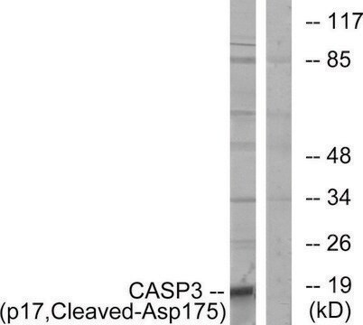
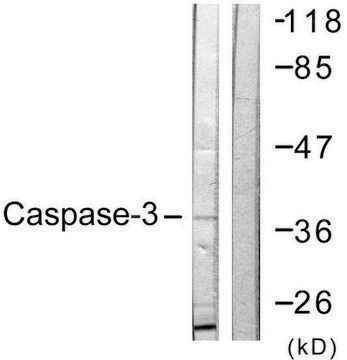
Western Blotting Analysis: 1μg/mL of this antibody detects Caspase-3 in A431 cell lysate.

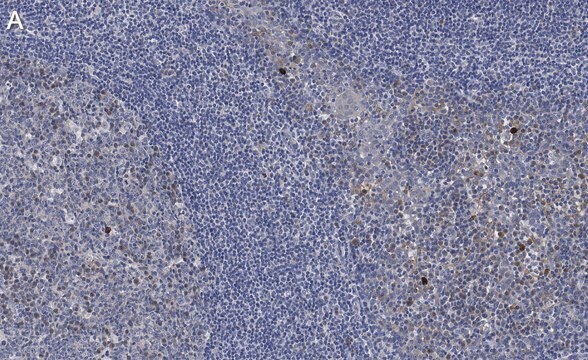
Immunohistochemistry (Paraffin) Analysis: A 1:250 dilution of this antibody detected Caspase-3 in Human tonsil tissue sections.

Minőség

routinely evaluated by immunoblot on RIPA lysates from non-stimulated human A431 cells, mouse 3T3/A31 or rat PC12 cells

Cél megnevezése

32 kDa

Kapcsolódás

Replaces: 04-1090; 04-439

Fizikai forma

Format: Purified
Protein A purified IgG in of 0.1M Tris-glycine, pH 7.4, 0.15M NaCl,and 0.05% sodium azide.

Tárolás és stabilitás

Stable for 2 years at 2-8°C from date of shipment. For maximum recovery of product, centrifuge the original vial prior to removing the cap.

Analízis megjegyzés

Control
Positive Antigen Control: Catalog #12-301, non-stimulated A431 cell lysate. Add 2.5µL of 2-mercaptoethanol/100µL of lysate and boil for 5 minutes to reduce the preparation. Load 20µg of reduced lysate per lane for mingels.

Jogi információk

UPSTATE is a registered trademark of Merck KGaA, Darmstadt, Germany

Nem találja a megfelelő terméket?  

Próbálja ki a Termékválasztó eszköz. eszközt

Tárolási osztály kódja

10 - Combustible liquids

WGK

WGK 1


Analitikai tanúsítványok (COA)

Analitikai tanúsítványok (COA) keresése a termék sarzs-/tételszámának megadásával. A sarzs- és tételszámok a termék címkéjén találhatók, a „Lot” vagy „Batch” szavak után.

Már rendelkezik ezzel a termékkel?

Az Ön által nemrégiben megvásárolt termékekre vonatkozó dokumentumokat a Dokumentumtárban találja.

Dokumentumtár megtekintése

Antiandrogen-induced cell death in LNCaP human prostate cancer cells.
Lee, EC; Zhan, P; Schallhom, R; Packman, K; Tenniswood, M
Cell Death and Differentiation null
Prisca Boisguérin et al.
Cardiovascular research, 116(3), 633-644 (2019-05-31)
Regulated cell death is a main contributor of myocardial ischaemia-reperfusion (IR) injury during acute myocardial infarction. In this context, targeting apoptosis could be a potent therapeutical strategy. In a previous study, we showed that DAXX (death-associated protein) was essential for
A caspase cascade regulating developmental axon degeneration.
Simon, DJ; Weimer, RM; McLaughlin, T; Kallop, D; Stanger, K; Yang, J; O'Leary et al.
The Journal of Neuroscience null
M Leist et al.
Biochemical and biophysical research communications, 258(1), 215-221 (1999-05-01)
The endogenous mediator nitric oxide (NO) blocked apoptosis of Jurkat cells elicited by staurosporine, anti-CD95 or chemotherapeutics, and switched death to necrosis. The switch in the mode of cell death was dependent on the ATP loss elicited by NO. This
Ying Wang et al.
Neuroreport, 23(18), 1052-1058 (2012-11-01)
Necrosis and apoptosis are well established as two primary cell death pathways. Mixed neuroglial cultures are commonly used to study cell death mechanisms in neural cells. However, the ages of these cultures vary across studies and little attention has been

Tudóscsoportunk valamennyi kutatási területen rendelkezik tapasztalattal, beleértve az élettudományt, az anyagtudományt, a kémiai szintézist, a kromatográfiát, az analitikát és még sok más területet.

Lépjen kapcsolatba a szaktanácsadással