Ugrás a tartalomra
Merck
Összes fotó(3)

Fontos dokumentumok

06-427

Sigma-Aldrich

Anti-Phosphotyrosine Antibody

Upstate®, from rabbit

Bejelentkezésa Szervezeti és Szerződéses árazás megtekintéséhez


About This Item

UNSPSC kód:
12352203
eCl@ss:
32160702
NACRES:
NA.41
klón:
polyclonal
application:
IP
WB
faj reaktivitás:
human
technika/technikák:
immunoprecipitation (IP): suitable
western blot: suitable
citations:
34

biológiai forrás

rabbit

Minőségi szint

antitest forma

affinity purified immunoglobulin

antitest terméktípus

primary antibodies

klón

polyclonal

tisztítva

affinity chromatography

faj reaktivitás

human

faj reaktivitás (homológia által előrejelzett)

all (based on 100% sequence homology)

gyártó/kereskedő neve

Upstate®

technika/technikák

immunoprecipitation (IP): suitable
western blot: suitable

kiszállítva

wet ice

célzott transzláció utáni módosítás

acetylation (test)

Géninformáció

human ... PID1(55022)

Általános leírás

The phosphorylation of specific tyrosine residues has been shown to be a primary mechanism of signal transduction during normal mitogenesis, cell cycle progression and oncogenic transformation, its role in other areas such as differentiation and gap junction communication, is a matter of active and ongoing research. Antibodies that specifically recognize phosphorylated tyrosine residues have proved to be invaluable to the study of tyrosine phosphorylated proteins and the biochemical pathways in which they function.

Egyediség

The immunoreactivity of the antibody is totally inhibited by the use of 100 mM phenyl phosphate, a phosphotyrosine analog. The phosphotyrosine antibody is purified by immunoaffinity chromatography using either a dual phospho-peptide gel or a BSA-phosphotyrosine gel. All of the phosphotyrosine immunoreactivity present in the antisera is immunoadsorbed whether the antibody is purified by either gel indicating that the antibody is not sequence-specific but specific for phosphotyrosine residues.

Immunogén

In order to produce broad spectrum polyclonal phosphotyrosine antibodies, rabbits were immunized with three phosphorylated immunogens: (1) phosphotyrosine covalently linked to KLH; (2) the c-Src carboxyl terminal regulatory phosphopeptide (TS-T-E-P-Q-pY-Q-P-G-E-N-L; Catalog # 12-218) covalently linked to KLH, and; (3) a phosphopeptide associated with high tyrosine kinase activity in human lymphocytes (R-R-L-I-E-D-A-E-pY-A-A-R-G; Catalog # 12-217) covalently linked to KLH. Both of the phosphopeptide haptens serve as strong substrates for tyrosine phosphatases and are part of the two colorimetric tyrosine phosphatase kits provided by Upstate, Inc. (Catalog # 17-125, 17-126).

Alkalmazás

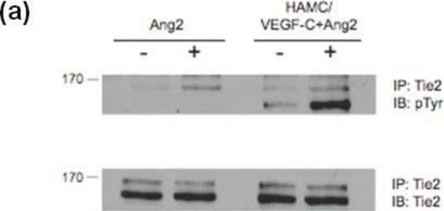
Anti-Phosphotyrosine Antibody detects level of Phosphotyrosine & has been published & validated for use in IP & WB.
Research Category
Signaling
Research Sub Category
General Post-translation Modification

Signaling Neuroscience
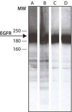
Western Blotting Analysis: A 1:500 dilution from a representative lot detected Phosphotyrosine in 10 µg of EGF treated A431 with Lambda Phosphatase tissue lysate.
Immunoprecipitation Analysis: 5 µL from a representative lot immunoprecipitated Phosphotyrosine in 0.5 mg of EGF treated A431 cell lysate

Minőség

Evaluated by Western Blotting in EGF treated A431 cell lysate.

Western Blotting Analysis: A 1:500 dilution of this antibody detected Phosphotyrosine in 10 µg of EGF treated A431 cell lysate.

Cél megnevezése

Varies

Fizikai forma

Affinity purified
Purified rabbit polyclonal in buffer containing 0.1 M Tris-Glycine (pH 7.4), 150 mM NaCl with 0.05% sodium azide.

Tárolás és stabilitás

Stable for 1 year at 2-8°C from date of receipt.

Analízis megjegyzés

Control
Positive Antigen Control: Catalog #12-302, EGF-stimulated A431 cell lysate. Add 2.5µL of 2-mercaptoethanol/100µL of lysate and boil for 5 minutes to reduce the preparation. Load 20µg of reduced lysate per lane for minigels.

Egyéb megjegyzések

Concentration: Please refer to the Certificate of Analysis for the lot-specific concentration.

Jogi információk

UPSTATE is a registered trademark of Merck KGaA, Darmstadt, Germany

Jogi nyilatkozat

Unless otherwise stated in our catalog or other company documentation accompanying the product(s), our products are intended for research use only and are not to be used for any other purpose, which includes but is not limited to, unauthorized commercial uses, in vitro diagnostic uses, ex vivo or in vivo therapeutic uses or any type of consumption or application to humans or animals.

Nem találja a megfelelő terméket?  

Próbálja ki a Termékválasztó eszköz. eszközt

WGK

WGK 1

Lobbanási pont (F)

does not flash

Lobbanási pont (C)

does not flash


Analitikai tanúsítványok (COA)

Analitikai tanúsítványok (COA) keresése a termék sarzs-/tételszámának megadásával. A sarzs- és tételszámok a termék címkéjén találhatók, a „Lot” vagy „Batch” szavak után.

Már rendelkezik ezzel a termékkel?

Az Ön által nemrégiben megvásárolt termékekre vonatkozó dokumentumokat a Dokumentumtárban találja.

Dokumentumtár megtekintése

In vivo insulin signaling through PI3-kinase is impaired in skeletal muscle of adult rat offspring exposed to ethanol in utero.
Chen, L; Yao, XH; Nyomba, BL
Journal of Applied Physiology (1985)
Human papillomavirus type 6b virus-like particles are able to activate the Ras-MAP kinase pathway and induce cell proliferation
Payne, E., et al
Journal of virology, 75, 4150-4157 (2001)
Selective activation of T cell kinase p56lck by Herpesvirus saimiri protein tip
Wiese, N., et al
The Journal of Biological Chemistry, 271, 847-852 (1996)
Yuan Gao et al.
PloS one, 8(6), e68105-e68105 (2013-07-11)
Chronic growth hormone (GH) therapy has been shown to cause insulin resistance, but the mechanism remains unknown. PTEN, a tumor suppressor gene, is a major negative regulator of insulin signaling. In this study, we explored the effect of chronic GH
Pawan K Vohra et al.
Biochimica et biophysica acta, 1833(12), 2953-2960 (2013-08-01)
Exogenous brain-derived neurotrophic factor (BDNF) enhances Ca(2+) signaling and cell proliferation in human airway smooth muscle (ASM), especially with inflammation. Human ASM also expresses BDNF, raising the potential for autocrine/paracrine effects. The mechanisms by which ASM BDNF secretion occurs are

Tudóscsoportunk valamennyi kutatási területen rendelkezik tapasztalattal, beleértve az élettudományt, az anyagtudományt, a kémiai szintézist, a kromatográfiát, az analitikát és még sok más területet.

Lépjen kapcsolatba a szaktanácsadással