Ugrás a tartalomra
Merck
Összes fotó(5)

Fontos dokumentumok

C2562

Sigma-Aldrich

Monoclonal Anti-Cytokeratin, pan (Mixture) antibody produced in mouse

clone C-11+PCK-26+CY-90+KS-1A3+M20+A53-B/A2, ascites fluid

Szinonimák:

Monoclonal Anti-Cytokeratin, pan (mixture), Panck Antibody, Panck Antibody - Monoclonal Anti-Cytokeratin, pan (Mixture) antibody produced in mouse

Bejelentkezésa Szervezeti és Szerződéses árazás megtekintéséhez


About This Item

MDL-szám:
UNSPSC kód:
12352203
NACRES:
NA.41
konjugátum:
unconjugated
application:
IF
IHC (f)
IHC (p)
WB
klón:
C-11+PCK-26+CY-90+KS-1A3+M20+A53-B/A2, monoclonal
faj reaktivitás:
wide range
citations:
162
technika/technikák:
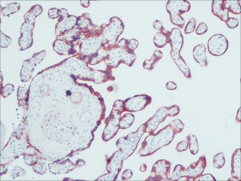
immunohistochemistry (formalin-fixed, paraffin-embedded sections): suitable using protease-digested sections of human or animal tissues
immunohistochemistry (frozen sections): suitable
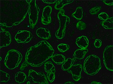
indirect immunofluorescence: 1:100 using protease-digested, formalin-fixed, paraffin-embedded sections of human or animal tissues
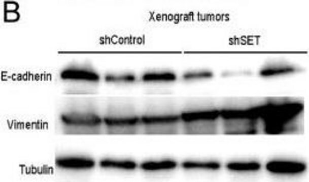
western blot: suitable

biológiai forrás

mouse

Minőségi szint

konjugátum

unconjugated

antitest forma

ascites fluid

antitest terméktípus

primary antibodies

klón

C-11+PCK-26+CY-90+KS-1A3+M20+A53-B/A2, monoclonal

tartalmaz

7% horse serum and 15 mM sodium azide as preservative

faj reaktivitás

wide range

technika/technikák

immunohistochemistry (formalin-fixed, paraffin-embedded sections): suitable using protease-digested sections of human or animal tissues
immunohistochemistry (frozen sections): suitable
indirect immunofluorescence: 1:100 using protease-digested, formalin-fixed, paraffin-embedded sections of human or animal tissues
western blot: suitable

izotípus

IgG1/IgG2a

kiszállítva

dry ice

tárolási hőmérséklet

−20°C

célzott transzláció utáni módosítás

unmodified

Looking for similar products? Látogasson el ide Útmutató a termékösszehasonlításhoz

Általános leírás

Monoclonal Anti-Cytokeratin, pan (mouse IgG1 and IgG2a isotypes) is a mixture of monoclonal antibodies from the following clones: C-11, PCK-26, CY-90, KS1A3, M20, and A53-B/A2. Cytokeratins, a group comprising at least 29 different proteins, are characteristic of epithelial and trichocytic cells. Cytokeratins 1 (68 kDa), 4 (59 kDa), 5 (58 kDa), 8 (52 kDa), and 6 (56 kDa) are members of the type II neutral-to-basic subfamily expressed in cornified epithelia, non-cornified stratified squamous epithelia, stratified epithelia, simple epithelia and in tissues with natural or pathological turnover respectively. Cytokeratins 10 (56 kDa), 13 (54 kDa, and 18 (45 kDa) are members of the type I acidic subfamily expressed in cornified epithelia, non-cornified stratified squamous epithelia and simple epithelial cells respectively. Cytokeratin peptide 19 (40 kDa) is a type I keratin which can be expressed in both simple and in broad cells of stratifying epithelia at specific sites.

Egyediség

This is a mixture of monoclonal anti-cytokeratin antibodies. It recognizes human cytokeratins 1,4,5,6,8,10,13,18, and 19. It is a broad spectrum reagent, which reacts specifically with a wide variety of normal, reactive, and neoplastic epithelial tissues. The antibody mixture reacts with simple, cornifying and non-cornifying squamous epithelia and pseudostratified epithelia. It does not react with non-epithelial normal human tissues. Increased staining intensity is seen following proteolytic treatment (protease unmasking).

Immunogén

mixture of several monoclonal cytokeratin clones.

Alkalmazás

Monoclonal Anti-Cytokeratin, pan (Mixture) antibody produced in mouse is suitable for the following applications:
  • Immunohistochemistry (formalin-fixed, paraffin-embedded sections) using protease-digested sections of human or animal tissues.
  • Immunohistochemistry (frozen sections).
  • Indirect immunofluorescence (at a working dilution of 1:100 using protease-digested, formalin-fixed, paraffin-embedded sections of human or animal tissues).
  • Immunocytochemical labeling (immunofluorescence ) of cells.
  • Western blotting.

Biokémiai/fiziológiai hatások

Cytokeratins are proteins of keratin-containing intermediate filaments that provide mechanical support and other additional functions in the epithelial cells. It is found in the intracytoplasmic cytoskeleton of epithelial tissue. Epithelial tissue expresses cytokeratin subunits in a specific and stable pattern. Cytokeratins along with vimentin are involved in cell proliferation, migration and differentiation of preodontoblasts and preameloblasts. The intermediate-sized filaments are abundant in human endothelial cells and are mostly of vimentin type.

Jogi nyilatkozat

Unless otherwise stated in our catalog or other company documentation accompanying the product(s), our products are intended for research use only and are not to be used for any other purpose, which includes but is not limited to, unauthorized commercial uses, in vitro diagnostic uses, ex vivo or in vivo therapeutic uses or any type of consumption or application to humans or animals.

Nem találja a megfelelő terméket?  

Próbálja ki a Termékválasztó eszköz. eszközt

Tárolási osztály kódja

10 - Combustible liquids

WGK

WGK 3


Válasszon a legfrissebb verziók közül:

Analitikai tanúsítványok (COA)

Lot/Batch Number

Nem találja a megfelelő verziót?

Ha egy adott verzióra van szüksége, a tétel- vagy cikkszám alapján rákereshet egy adott tanúsítványra.

Már rendelkezik ezzel a termékkel?

Az Ön által nemrégiben megvásárolt termékekre vonatkozó dokumentumokat a Dokumentumtárban találja.

Dokumentumtár megtekintése

J M Pérez-Pomares et al.
Developmental dynamics : an official publication of the American Association of Anatomists, 210(2), 96-105 (1997-10-23)
A study about the hypothetical contribution of the epicardial cells to the subepicardial mesenchyme was carried out in Syrian hamster embryos of 9-12 days post coitum (dpc) and chick embryos of 3-5 days of incubation. In the epicardium and subepicardium
Structure and functions of keratin proteins in simple, stratified, keratinized and cornified epithelia
Bragulla HH and Homberger DG
Journal of Anatomy, 214(4), 516-559 (2009)
Emmanuel Letavernier et al.
Nephrology, dialysis, transplantation : official publication of the European Dialysis and Transplant Association - European Renal Association, 24(2), 630-638 (2008-10-18)
The specific mTor inhibitor sirolimus has been implicated in the pathogenesis of renal glomerular lesions and nephrotic syndrome appearance after transplantation. Podocyte injury and focal segmental glomerulosclerosis have been related to sirolimus therapy in some patients but the pathways underlying
R Muñoz-Chápuli et al.
Differentiation; research in biological diversity, 64(3), 133-141 (1999-05-11)
The existence of the hemangioblast, a common progenitor of the endothelial and hematopoietic cell lineages, was proposed at the beginning of the century. Although recent findings seem to confirm its existence, it is still unknown when and how the hemangioblasts
Marta Gabasa et al.
PloS one, 8(6), e65445-e65445 (2013-06-12)
Prostaglandin E2 (PGE2), the main metabolite of cyclooxygenase (COX), is a well-known anti-fibrotic agent. Moreover, myofibroblasts expressing α-smooth muscle actin (α-SMA), fibroblast expansion and epithelial-mesenchymal transition (EMT) are critical to the pathogenesis of idiopathic pulmonary fibrosis (IPF). Our aim was

Tudóscsoportunk valamennyi kutatási területen rendelkezik tapasztalattal, beleértve az élettudományt, az anyagtudományt, a kémiai szintézist, a kromatográfiát, az analitikát és még sok más területet.

Lépjen kapcsolatba a szaktanácsadással