05-915
Anti-N-Cadherin Antibody, clone 13A9
culture supernatant, clone 13A9, Upstate®
Szinonimák:
Cadherin-2, CD325, CDw325, N-cadherin, Neural cadherin
About This Item
IHC
IP
WB
immunohistochemistry: suitable
immunoprecipitation (IP): suitable
western blot: suitable
Javasolt termékek
biológiai forrás
mouse
Minőségi szint
antitest forma
culture supernatant
antitest terméktípus
primary antibodies
klón
13A9, monoclonal
faj reaktivitás
human
kiszerelés
antibody small pack of 25 μL
gyártó/kereskedő neve
Upstate®
technika/technikák
immunocytochemistry: suitable
immunohistochemistry: suitable
immunoprecipitation (IP): suitable
western blot: suitable
NCBI elérési szám
UniProt elérési szám
kiszállítva
ambient
célzott transzláció utáni módosítás
unmodified
Géninformáció
human ... CDH2(1000)
Related Categories
Általános leírás
Egyediség
Immunogén
Alkalmazás
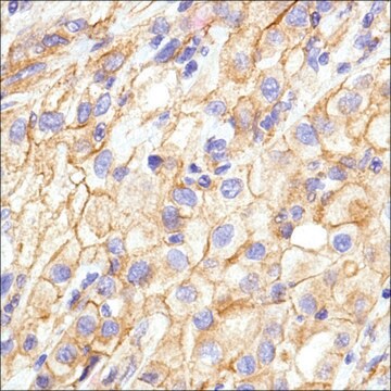
Immunohistochemistry Analysis: A representative lot detected strong N-cadherin immunoreactivity in paraffin-embedded rectal cancer (RC) tissues with positive regional lymph node metastasis (RLNM) status, while only weak N-cadherin immunoreactivity was detected in RC with negative RLNM, and no N-cadherin staining was seen in normal colorectal epithelium (Fan, X.J., et al. (2012). Br. J. Cancer. 106(11):1735-1741).
Immunohistochemistry Analysis: A representative lot detected N-cadherin immunoreactivity in formalin-fixed, paraffin-embedded hepatocellular carcinoma (HCC) tissue sections. A significant inverse correlation was found between RUNX3 and N-cadherin expression levels (Tanaka, S., et al. (2012). Int. J. Cancer. 131(11):2537-2546).
Western Blotting Analysis: A representative lot detected an upregulated N-cadherin expression in CCL185 carcinoma cells following transient Epstein-Barr virus (EBV) infection. The EMT-like phenotype remained even after viral loss by culture selection pressure withdrawal (Queen, K.J., et al. (2013). Int. J. Cancer. 132(9):2076-2086).
Western Blotting Analysis: A representative lot detected N-cadherin in Hep3B, Huh7, HLF and SK-Hep1 human hepatocellular carcinoma (HCC) cell lysates (Tanaka, S., et al. (2012). Int. J. Cancer. 131(11):2537-2546).
Western Blotting Analysis: A representative lot detected both the unprocessed (pro-) and processed (mature) forms of N-cadherin in HeLa cell lysate (Wahl, J.K. 3rd., et al. (2003). J. Biol. Chem. 278(19):17269-17276).
Western Blotting Analysis: A representative lot detected N-cadherin in WI-38 human fibroblast lysate, but not in JAr human placental choriocarcinoma cell lysate (Knudsen, K.A., et al. (1995). J. Cell Biol. 130(1):67-77).
Immunocytochemistry Analysis: A representative lot detected N-cadherin immunoreactivity localized primarily at the cell-cell borders by fluorescent immunocytochemistry staining of 1% paraformaldehyde-fixed, methanol-permeabilized HeLa cells (Wahl, J.K. 3rd., et al. (2003). J. Biol. Chem. 278(19):17269-17276).
Immunocytochemistry Analysis: A representative lot detected N-cadherin immunoreactivity colocalized with those of alpha- and beta-catenin by dual fluorescent immunocytochemistry staining of fixed WI-38 human fibroblasts (Knudsen, K.A., et al. (1995). J. Cell Biol. 130(1):67-77).
Immunoprecipitation Analysis: Representative lots co-immunoprecipitated alpha-catenin, beta-catenin, and plakoglobin with N-cadherin from WI-38 human fibroblast and HeLa cell lysates (Wahl, J.K. 3rd., et al. (2003). J. Biol. Chem. 278(19):17269-17276; Knudsen, K.A., et al. (1995). J. Cell Biol. 130(1):67-77).
Cell Structure
Adhesion (CAMs)
Minőség
Western Blotting Analysis: A 1:1000-5000 dilution of this hybridoma culture supernatant detected N-cadherin in HeLa cell lysate.
Cél megnevezése
Kapcsolódás
Fizikai forma
Tárolás és stabilitás
Analízis megjegyzés
HeLa cell lysate
Jogi információk
Jogi nyilatkozat
Nem találja a megfelelő terméket?
Próbálja ki a Termékválasztó eszköz. eszközt
javasolt
Analitikai tanúsítványok (COA)
Analitikai tanúsítványok (COA) keresése a termék sarzs-/tételszámának megadásával. A sarzs- és tételszámok a termék címkéjén találhatók, a „Lot” vagy „Batch” szavak után.
Már rendelkezik ezzel a termékkel?
Az Ön által nemrégiben megvásárolt termékekre vonatkozó dokumentumokat a Dokumentumtárban találja.
Cikkek
Humán iPSC neurális differenciálódási közegek és protokollok, amelyeket neurális őssejtek, neuronok és gliasejt-típusok létrehozására használnak.
Human iPSC neural differentiation media and protocols used to generate neural stem cells, neurons and glial cell types.
Tudóscsoportunk valamennyi kutatási területen rendelkezik tapasztalattal, beleértve az élettudományt, az anyagtudományt, a kémiai szintézist, a kromatográfiát, az analitikát és még sok más területet.
Lépjen kapcsolatba a szaktanácsadással


