
MAB4234
Anti-Wilms′ Tumor Antibody, NT, clone 6F-H2
clone 6F-H2, Chemicon®, from mouse
Szinonimák:
WT1
About This Item
Javasolt termékek


biológiai forrás
mouse
Minőségi szint
antitest forma
purified immunoglobulin
antitest terméktípus
primary antibodies
klón
6F-H2, monoclonal
faj reaktivitás
human, mouse
gyártó/kereskedő neve
Chemicon®
technika/technikák
immunocytochemistry: suitable
immunofluorescence: suitable
immunohistochemistry: suitable (paraffin)
immunoprecipitation (IP): suitable
western blot: suitable
izotípus
IgG1κ
NCBI elérési szám
UniProt elérési szám
kiszállítva
wet ice
célzott transzláció utáni módosítás
unmodified
Géninformáció
human ... WT1(7490)
Általános leírás
Egyediség
Immunogén
Alkalmazás
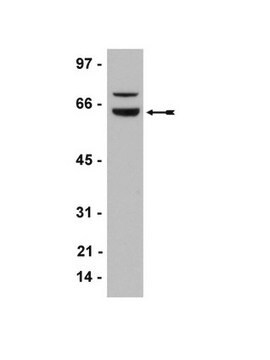
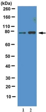
Western Blotting Analysis: A representative lot detected Wilms tumor protein WT1 in the CRE-binding protein/CBP immunoprecipitate obtained from the lysate of a T-SV40 immortalized human glomerular epithelial cell (HGEC) line (Drossopoulou, G.I., et al. (2009). Am. J. Physiol. Renal Physiol. 297(3):F594-F603).
Western Blotting Analysis: A representative lot detected Wilms tumor protein WT1 in lysates from mouse E15.5 embryonic kidney and human melanoma cell lines A375, SK-MEL-28, and WM-266-4 (Wagner, N., et al. (2008). Pflugers Arch. Eur. J. Physiol. 455(5):839-847).
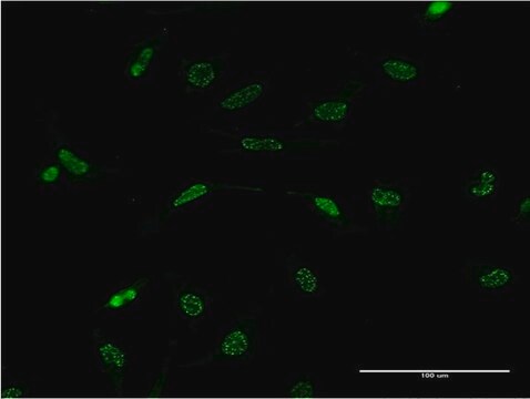
Immunocytochemistry Analysis: A representative lot immunostained the nucleus of methanol-fixed human melanoma A375 cells by fluorescent immunocytochemistry (Wagner, N., et al. (2008). Pflugers Arch. Eur. J. Physiol. 455(5):839-847).
Immunofluorescence Analysis: A representative lot immunostained the PCNA-positive nuclei of proliferating cells in formalin-fixed, paraffin-embedded human melanoma tissue sections by fluorescent immunohistochemistry (Wagner, N., et al. (2008). Pflugers Arch. Eur. J. Physiol. 455(5):839-847).
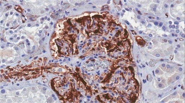
Immunohistochemistry Analysis: A representative lot immunostained glomeruli in formalin-fixed, paraffin-embedded normal human kidney and Wilms′ tumor sections (Wagner, N., et al. (2008). Pediatr. Nephrol. 23(9):1445-1453).
Immunohistochemistry Analysis: A representative lot detected vascular WT1 expression in 95% of 113 paraffin-embedded tumour tissues of various types. In most cases, nuclear WT1 staining of endothelial cells was seen (Wagner, N., et al. (2008). Oncogene. 27(26):3662-3672).
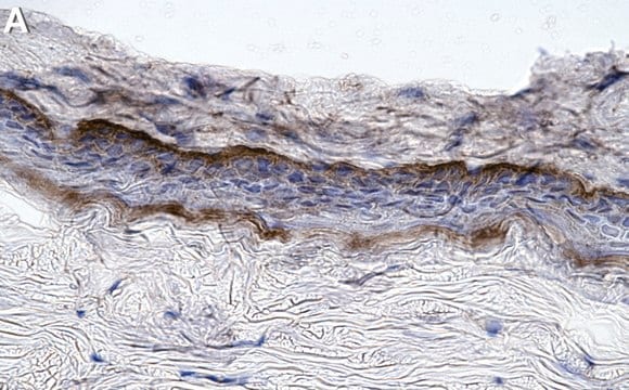
Immunohistochemistry Analysis: A representative lot immunostained the nucleus of perifollicular fibroblasts at the hair follicle in formalin-fixed, paraffin-embedded normal human skin sections. Most common melanocytic nevi do not express WT1, whereas Spitz nevi and dysplastic nevi show cytoplasmic WT1 staining. (Wagner, N., et al. (2008). Pflugers Arch. Eur. J. Physiol. 455(5):839-847).
Epigenetics & Nuclear Function
Transcription Factors
Cél megnevezése
Fizikai forma
Tárolás és stabilitás
Egyéb megjegyzések
Jogi információk
Jogi nyilatkozat
Nem találja a megfelelő terméket?
Próbálja ki a Termékválasztó eszköz. eszközt
javasolt
Tárolási osztály kódja
10 - Combustible liquids
WGK
WGK 2
Lobbanási pont (F)
Not applicable
Lobbanási pont (C)
Not applicable
Analitikai tanúsítványok (COA)
Analitikai tanúsítványok (COA) keresése a termék sarzs-/tételszámának megadásával. A sarzs- és tételszámok a termék címkéjén találhatók, a „Lot” vagy „Batch” szavak után.
Már rendelkezik ezzel a termékkel?
Az Ön által nemrégiben megvásárolt termékekre vonatkozó dokumentumokat a Dokumentumtárban találja.
Tudóscsoportunk valamennyi kutatási területen rendelkezik tapasztalattal, beleértve az élettudományt, az anyagtudományt, a kémiai szintézist, a kromatográfiát, az analitikát és még sok más területet.
Lépjen kapcsolatba a szaktanácsadással