Ugrás a tartalomra
Merck
Összes fotó(2)

Fontos dokumentumok

AB5407

Sigma-Aldrich

Anti-Opsin Antibody, blue

Chemicon®, from rabbit

Szinonimák:

Anti-BCP, Anti-BOP

Bejelentkezésa Szervezeti és Szerződéses árazás megtekintéséhez


About This Item

UNSPSC kód:
12352203
eCl@ss:
32160702
NACRES:
NA.41
klón:
polyclonal
application:
IHC
faj reaktivitás:
monkey, human, mouse
technika/technikák:
immunohistochemistry: suitable (paraffin)
citations:
147

biológiai forrás

rabbit

Minőségi szint

antitest forma

purified immunoglobulin

antitest terméktípus

primary antibodies

klón

polyclonal

faj reaktivitás

monkey, human, mouse

gyártó/kereskedő neve

Chemicon®

technika/technikák

immunohistochemistry: suitable (paraffin)

NCBI elérési szám

UniProt elérési szám

kiszállítva

wet ice

célzott transzláció utáni módosítás

unmodified

Géninformáció

human ... OPN1LW(5956)

Általános leírás

The full range of color discrimination in humans is based on the presence and function of three cone photoreceptor mechanisms. Each cone type possesses a photo-sensitive pigment-protein complex consisting of 11-cis retinal and a unique opsin protein, which gives sensitivity in the short (S cone, peak sensitivity about 420nm), middle (M cone, peak sensitivity about 530nm with polymorphism; Winderckx et al., 1993; Neitz & Neitz, 1998), and long (L cone, peak sensitivity about 560nm with polymorphism; Neitz & Jacobs, 1990) wavelengths of the light spectrum. All three opsins are transmembrane proteins with seven membrane-spanning regions. Genes for the three types of cone opsins and the rod photoreceptor rhodopsin gene seem to be homologous with varying amounts of conservation. Strongest conservation is between the middle (green) and long (red) wavelength sensitive pigments on the X chromosome, suggesting a relatively recent duplication/divergence event (Nathans, 1989; Nathans et al., 1992). The S cone (blue) opsin is located on chromosome 7 and seems to have stronger conservation with rhodopsin. Cone photoreceptor distribution in humans is dominated by the M and L cone pigments.

Egyediség

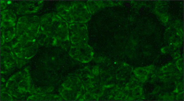
Recognizes Opsin, blue.

Immunogén

Epitope: blue
Recombinant human blue opsin.

Alkalmazás

Immunohistochemistry: 1:200-1:300 on formalin-fixed, paraffin-embedded mouse retina tissue. Antigen retrieval method recommend is HIER with steam heat; other fixation and retrieval methods are untested.

Optimal working dilutions must be determined by the end user.
Research Category
Neuroscience
Research Sub Category
Sensory & PNS
This Anti-Opsin Antibody, blue is validated for use in IH(P) for the detection of Opsin.

Fizikai forma

Format: Purified
Protein A purified
Purified immunoglobulinin PBS {0.02M phosphate, 0.25M NaCl, pH 7.6} with 0.1% sodium azide as a preservative

Tárolás és stabilitás

Maintain for 1 year at 2–8°C from date of shipment.

Analízis megjegyzés

Control
Retina

Jogi információk

CHEMICON is a registered trademark of Merck KGaA, Darmstadt, Germany

Jogi nyilatkozat

Unless otherwise stated in our catalog or other company documentation accompanying the product(s), our products are intended for research use only and are not to be used for any other purpose, which includes but is not limited to, unauthorized commercial uses, in vitro diagnostic uses, ex vivo or in vivo therapeutic uses or any type of consumption or application to humans or animals.

Nem találja a megfelelő terméket?  

Próbálja ki a Termékválasztó eszköz. eszközt

Tárolási osztály kódja

12 - Non Combustible Liquids

WGK

WGK 1

Lobbanási pont (F)

Not applicable

Lobbanási pont (C)

Not applicable


Analitikai tanúsítványok (COA)

Analitikai tanúsítványok (COA) keresése a termék sarzs-/tételszámának megadásával. A sarzs- és tételszámok a termék címkéjén találhatók, a „Lot” vagy „Batch” szavak után.

Már rendelkezik ezzel a termékkel?

Az Ön által nemrégiben megvásárolt termékekre vonatkozó dokumentumokat a Dokumentumtárban találja.

Dokumentumtár megtekintése

Photoreceptor coupling mediated by connexin36 in the primate retina.
O'Brien, JJ; Chen, X; Macleish, PR; O'Brien, J; Massey, SC
The Journal of Neuroscience null
Prasanthi Namburi et al.
American journal of human genetics, 99(3), 777-784 (2016-09-03)
Inherited retinal diseases (IRDs) are a diverse group of genetically and clinically heterogeneous retinal abnormalities. The present study was designed to identify genetic defects in individuals with an uncommon combination of autosomal recessive progressive cone-rod degeneration accompanied by sensorineural hearing
L P Morin et al.
Neuroscience, 199, 213-224 (2011-10-12)
Four studies were performed to further clarify the contribution of rod/cone and intrinsically photoreceptive retinal ganglion cells to measures of entrainment, dark preference, light-induced locomotor suppression and photosomnolence. Wild type (WT), retinally degenerate (rd/rd), and melanopsin-less (OPN4⁻/⁻) mouse strains were
Peter H Tang et al.
The Journal of neuroscience : the official journal of the Society for Neuroscience, 31(28), 10403-10411 (2011-07-15)
As cone photoreceptors mediate vision in bright light, their photopigments are bleached at a rapid rate and require substantial recycling of the chromophore 11-cis-retinal (RAL) for continued function. The retinal pigment epithelium (RPE) supplies 11-cis-RAL to both rod and cone
Xiaojie Ji et al.
Investigative ophthalmology & visual science, 57(3), 877-888 (2016-03-16)
Retinal detachments (RDs), a separation of the light-sensitive tissue of the retina from its supporting layers in the posterior eye, isolate retinal cells from their normal supply of nourishment and can lead to their deterioration and death. We identified a

Tudóscsoportunk valamennyi kutatási területen rendelkezik tapasztalattal, beleértve az élettudományt, az anyagtudományt, a kémiai szintézist, a kromatográfiát, az analitikát és még sok más területet.

Lépjen kapcsolatba a szaktanácsadással