Direkt zum Inhalt
Merck

T5530

Sigma-Aldrich

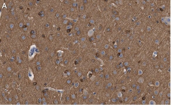
ANTI-TAU (MAPT) Antibody

mouse monoclonal, TAU-2

Anmeldenzur Ansicht organisationsspezifischer und vertraglich vereinbarter Preise

Größe auswählen

0.2 ML
€ 713,00

€ 713,00


Voraussichtliches Versanddatum21. Mai 2025

Für Ihr Target ist ein rekombinanter, konservierungsmittelfreier Antikörper verfügbar. Probieren Sie ZMS3420

Bulk-Bestellung anfordern

Größe auswählen

Ansicht ändern
0.2 ML
€ 713,00

About This Item

MDL-Nummer:
UNSPSC-Code:
12352203
NACRES:
NA.41

€ 713,00


Voraussichtliches Versanddatum21. Mai 2025

Für Ihr Target ist ein rekombinanter, konservierungsmittelfreier Antikörper verfügbar. Probieren Sie ZMS3420

Bulk-Bestellung anfordern

Produktbezeichnung

Monoclonal Anti-τ (Tau) antibody produced in mouse, clone TAU-2, ascites fluid

Biologische Quelle

mouse

Konjugat

unconjugated

Antikörperform

ascites fluid

Antikörper-Produkttyp

primary antibodies

Klon

TAU-2, monoclonal

Mol-Gew.

antigen 55-62 kDa

Enthält

15 mM sodium azide

Speziesreaktivität

monkey, bovine, chicken, human

Methode(n)

immunohistochemistry (formalin-fixed, paraffin-embedded sections): suitable
microarray: suitable
western blot: 1:1,000 using a fresh total bovine brain extract or an enriched microtubule protein preparation

Isotyp

IgG1

UniProt-Hinterlegungsnummer

Versandbedingung

dry ice

Lagertemp.

−20°C

Posttranslationale Modifikation Target

unmodified

Angaben zum Gen

human ... MAPT(4137)

Verwandte Kategorien

Allgemeine Beschreibung

Monoclonal Anti- τ (TAU) (mouse IgG1 isotype) is derived from the hybridoma produced by the fusion of mouse myeloma cells and splenocytes from an immunized mouse. τ (TAU) proteins are a part of microtubule associated proteins (MAPs). They are densely found in neurons and in trace amounts in non-neuronal cells. In brain six isoforms of τ (TAU) proteins are present.
The antibody reacts exclusively with the chemically heterogeneous τ in both the phosphorylated and non-phosphorylated form. The antibody does not react with other MAPs or with tubulin. In immunohistochemical staining, it localizes τ along microtubules in axons, somata, dendrites and astrocytes, and on ribosomes. The antibody may be used for staining of τ in Alzheimer neurofibrillary tangles in sections of human brain tissue.
The best known microtubule associated proteins (MAPs) which copurify with microtubules are MAP2 and Tau. These two proteins are heat stable and stimulate formation of the microtubule polymer from purified tubulin subunits. Tau is chemically heterogenous, however, limited protolysis has demonstrated that the different eletrophoretic species are closely related. Tau is immunologically distinct from the other MAPs, namely MAP1, MAP2 and MAP5. Localization studies have demonstrated that Tau is intimately associated with the filamentous structures which compose the neurofibrillary tangles as found in an Alzheimer′s disease brain.

Immunogen

bovine microtubule-associated proteins (MAPs)

Anwendung

Monoclonal Anti-τ (Tau) antibody has been used:
  • in immunohistology
  • in immunoblotting
  • in dot blot
  • in immunohistochemistry

Mouse monoclonal clone TAU-2 anti-Tau antibody maybe used to study microtubule associated proteins (MAP) expression and cytological localization in various tissue and cell lines, under different developmental and environmental circumstances.

Biochem./physiol. Wirkung

Monoclonal Anti- τ (TAU) is phosphatase independent; it will bind Tau proteins in either their phosphorylated or non-phosphorylated forms. It localizes Tau proteins along microtubules in axons, somata, dendrites, astrocytes and on ribosomes (polysomes). The best-known microtubule associated proteins (MAPs) which copurify with microtubules are MAP2 and Tau. These two proteins are heat stable and stimulate formation of the microtubule polymer from purified tubulin subunits. Tau is immunologically distinct from the other MAPs. Tau is intimately associated with the filamentous structures which compose the neurofibrillary tangles as found in an Alzheimer′s disease brain.

Haftungsausschluss

Unless otherwise stated in our catalog or other company documentation accompanying the product(s), our products are intended for research use only and are not to be used for any other purpose, which includes but is not limited to, unauthorized commercial uses, in vitro diagnostic uses, ex vivo or in vivo therapeutic uses or any type of consumption or application to humans or animals.

Sie haben nicht das passende Produkt gefunden?  

Probieren Sie unser Produkt-Auswahlhilfe. aus.

Lagerklassenschlüssel

10 - Combustible liquids

WGK

WGK 3

Flammpunkt (°F)

Not applicable

Flammpunkt (°C)

Not applicable


Hier finden Sie alle aktuellen Versionen:

Analysenzertifikate (COA)

Lot/Batch Number

Die passende Version wird nicht angezeigt?

Wenn Sie eine bestimmte Version benötigen, können Sie anhand der Lot- oder Chargennummer nach einem spezifischen Zertifikat suchen.

Besitzen Sie dieses Produkt bereits?

In der Dokumentenbibliothek finden Sie die Dokumentation zu den Produkten, die Sie kürzlich erworben haben.

Die Dokumentenbibliothek aufrufen

Chris R Guthrie et al.
Journal of molecular neuroscience : MN, 45(1), 32-41 (2011-02-23)
Lesions containing aggregated and hyperphosphorylated tau protein are characteristic of neurodegenerative tauopathies. We have developed a cellular model of pathological tau deposition and clearance by overexpressing wild type human tau in HEK293 cells. When proteasome activity is inhibited, HEK293/tau cells
Cortical limb myoclonus in pathologically proven progressive supranuclear palsy.
Sharon Kemp et al.
Movement disorders : official journal of the Movement Disorder Society, 28(13), 1804-1806 (2013-10-15)
Severity of gliosis in Pick?s disease and frontotemporal lobar degeneration: tau-positive glia differentiate these disorders
Schofield E, et al.
Brain, 126(4), 827-840 (2003)
R E Mrak et al.
Human pathology, 26(8), 816-823 (1995-08-01)
The roles of activated glia and of glial cytokines in the pathogenesis of Alzheimer's disease are reviewed. Interleukin-1 (IL-1), a microglia-derived acute phase cytokine, activates astrocytes and induces expression of the astrocyte-derived cytokine, S100 beta, which stimulates neurite growth (and
S Alladi et al.
Brain : a journal of neurology, 130(Pt 10), 2636-2645 (2007-09-28)
To determine the frequency of Alzheimer's disease (AD) pathology in patients presenting with progressive focal cortical syndromes, notably posterior cortical atrophy (PCA), corticobasal syndrome (CBS), behavioural variant frontotemporal dementia (bvFTD), progressive non-fluent aphasia (PNFA) (or a mixed aphasia) and semantic

Questions

Reviews

No rating value

Active Filters

Unser Team von Wissenschaftlern verfügt über Erfahrung in allen Forschungsbereichen einschließlich Life Science, Materialwissenschaften, chemischer Synthese, Chromatographie, Analytik und vielen mehr..

Setzen Sie sich mit dem technischen Dienst in Verbindung.