
おすすめの製品


由来生物
mouse
品質水準
抗体製品の状態
purified immunoglobulin
抗体製品タイプ
primary antibodies
クローン
2H4.2, monoclonal
交差性
human
テクニック
immunohistochemistry: suitable
western blot: suitable
アイソタイプ
IgG1κ
NCBIアクセッション番号
UniProtアクセッション番号
輸送温度
wet ice
ターゲットの翻訳後修飾
unmodified
遺伝子情報
human ... ATG13(9776)
詳細
The autophagy-related protein 13 (Atg13) plays an important role in the formation of autophagosomes. Autophagosomes are formed in response to nutrient deprivation and function as the transport vesicles for organelles, proteins, and protein complexes targeted for lysosomes that digest these cargos to produce energy and nutrients. Atg13 is activated by the mTOR pathway and forms a complex with the FIP200 protein. This complex is involved in enhancing the activity of the ULK1 kinase which is required for the formation of autophagosomes. Atg13/FIP200 faciliates the localization of ULK1 to pre-autophagosomes, and subsequently stabilizes ULK1. Autophagy is an important process in development, growth, and cell differentiation, and disruption of this process may contribute to cancer, aging, and neurodegenerative diseases.
免疫原
GST-tagged recombinant protein corresponding to human Atg13.
アプリケーション
Research Category
アポトーシス及び癌
アポトーシス及び癌
Research Sub Category
アポトーシス-追加
アポトーシス-追加
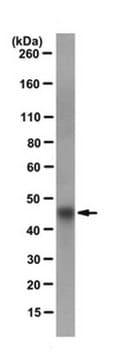
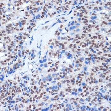
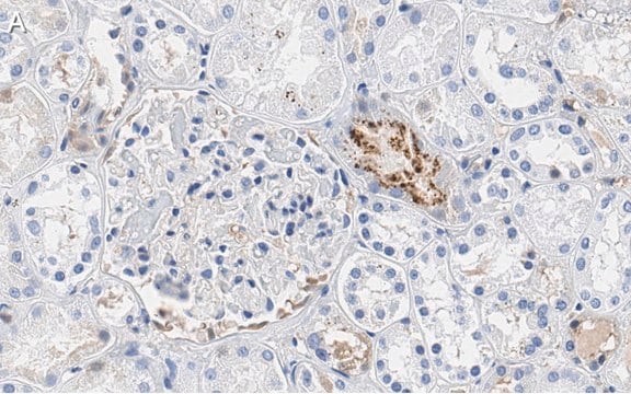
This Anti-Atg13 Antibody, clone 2H4.2 is validated for use in Western Blotting, IHC for the detection of Atg13.
Western Blot Analysis: A representative lot detected Atg13 in HeLa cell lysate.
Immunohistochemistry Analysis: A 1:50 dilution from a representative lot detected Atg13 in normal human substancia nigra tissues.
Immunohistochemistry Analysis: A 1:50 dilution from a representative lot detected Atg13 in normal human substancia nigra tissues.
品質
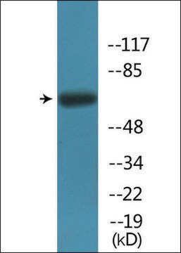
Evaluated by Western Blot in campthothecin treated HeLa cell lysate.
Western Blot Analysis: A 1:1,000 dilution of this antibody detected Atg13 in 10 µg of campthothecin treated HeLa cell lysate.
Western Blot Analysis: A 1:1,000 dilution of this antibody detected Atg13 in 10 µg of campthothecin treated HeLa cell lysate.
ターゲットの説明
~60 kDa observed
物理的形状
Protein G Purified
Format: Purified
Purified mouse monoclonal IgG1κ in buffer containing 0.1 M Tris-Glycine (pH 7.4), 150 mM NaCl with 0.05% sodium azide.
保管および安定性
Stable for 1 year at 2-8°C from date of receipt.
アナリシスノート
Control
Campthothecin treated HeLa cell lysate.
Campthothecin treated HeLa cell lysate.
免責事項
Unless otherwise stated in our catalog or other company documentation accompanying the product(s), our products are intended for research use only and are not to be used for any other purpose, which includes but is not limited to, unauthorized commercial uses, in vitro diagnostic uses, ex vivo or in vivo therapeutic uses or any type of consumption or application to humans or animals.
適切な製品が見つかりませんか。
製品選択ツール.をお試しください
保管分類コード
12 - Non Combustible Liquids
WGK
WGK 1
引火点(°F)
Not applicable
引火点(℃)
Not applicable
適用法令
試験研究用途を考慮した関連法令を主に挙げております。化学物質以外については、一部の情報のみ提供しています。 製品を安全かつ合法的に使用することは、使用者の義務です。最新情報により修正される場合があります。WEBの反映には時間を要することがあるため、適宜SDSをご参照ください。
Jan Code
MABC46:
試験成績書(COA)
製品のロット番号・バッチ番号を入力して、試験成績書(COA) を検索できます。ロット番号・バッチ番号は、製品ラベルに「Lot」または「Batch」に続いて記載されています。
Yo-Hei Yamamoto et al.
The Journal of cell biology, 219(8) (2020-06-04)
In macroautophagy, membrane structures called autophagosomes engulf substrates and deliver them for lysosomal degradation. Autophagosomes enwrap a variety of targets with diverse sizes, from portions of cytosol to larger organelles. However, the mechanism by which autophagosome size is controlled remains
Correction: ERdj8 governs the size of autophagosomes during the formation process.
Yo-Hei Yamamoto et al.
The Journal of cell biology, 220(9) (2021-08-07)
Ubiquitination Is a Novel Post-Translational Modification of VMP1 in Autophagy of Human Tumor Cells.
Felipe J Renna et al.
International journal of molecular sciences, 24(16) (2023-08-26)
Autophagy is a tightly regulated catabolic process involved in the degradation and recycling of proteins and organelles. Ubiquitination plays an important role in the regulation of autophagy. Vacuole Membrane Protein 1 (VMP1) is an essential autophagy protein. The expression of
Naoki Tamura et al.
Molecular and cellular biology, 39(16) (2019-06-05)
Autophagy is considered an adaptive mechanism against hyperosmotic stress. Although the process has been reported to be triggered by the inhibition of mTORC1, the precise downstream mechanisms remain elusive. Here, we demonstrate that hyperosmotic-stress-induced autophagy is different from conventional macroautophagy
Marianna Carinci et al.
The EMBO journal, 40(10), e103563-e103563 (2021-05-02)
The early secretory pathway and autophagy are two essential and evolutionarily conserved endomembrane processes that are finely interlinked. Although growing evidence suggests that intracellular trafficking is important for autophagosome biogenesis, the molecular regulatory network involved is still not fully defined.
アクティブなフィルタ
ライフサイエンス、有機合成、材料科学、クロマトグラフィー、分析など、あらゆる分野の研究に経験のあるメンバーがおります。.
製品に関するお問い合わせはこちら(テクニカルサービス)