
現在、価格および在庫状況を閲覧できません。
おすすめの製品


由来生物
rat
品質水準
結合体
unconjugated
抗体製品の状態
purified antibody
抗体製品タイプ
primary antibodies
クローン
5A6, monoclonal
分子量
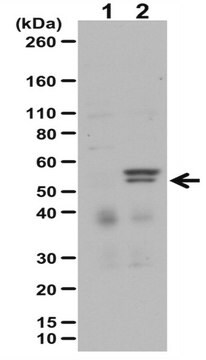
calculated mol wt 54.32 kDa
observed mol wt ~54 kDa
精製方法
using protein G
交差性
mouse
包装
antibody small pack of 100 μL
テクニック
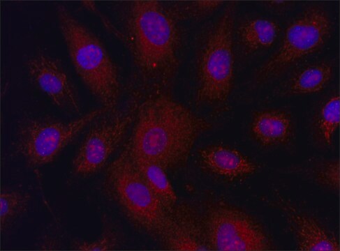
immunofluorescence: suitable
western blot: suitable
アイソタイプ
IgG2aλ
エピトープ配列
C-terminal
タンパク質IDアクセッション番号
UniProtアクセッション番号
輸送温度
ambient
ターゲットの翻訳後修飾
unmodified
遺伝子情報
mouse ... Mlkl(74568)
詳細
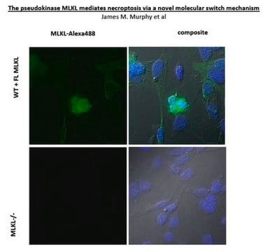
Mixed lineage kinase domain-like protein (UniProt: Q9D2Y4; also known as MLKL) is encoded by the Mlkl gene (Gene ID: 74568) in murine species. MLKL is a pseudokinase that is a terminal protein in the pro-inflammatory necroptotic cell death program. It plays a critical role in TNFa-induced necroptosis. It is highly expressed in thymus, colon, intestine, liver, spleen, and lung. Its expression is much lower in skeletal muscle, heart and kidney, and it is not detected in the brain. Although it contains a protein kinase domain (aa 192-456), it lacks several residues that are essential for protein kinase activity. It is activated following phosphorylation by RIPK3, which leads to its homotrimerization and localization to the plasma membrane where it binds to highly phosphorylated inositol (InsP6) and disrupts membrane integrity to cause necrotic cell death. Phosphorylation of MLKL at serine 345 is shown to be essential for necroptosis in murine cells. Its interaction with RIPK3 is species specific and mouse MLKL does not interact with human RIPK3. In contrast to human protein, mouse MLKL is not inhibited by necrosulfonamide, because at position 85 it contains a tryptophan residue instead of a cysteine. Under basal conditions, MLKL is shown to reside in small puncta that are distributed evenly throughout the cytoplasm, but it is detected in plasma membrane shortly after its phosphorylation. It contains two coiled coil regions (aa 61-81 and 138-229) and the second coiled coil region is responsible for its oligomerization. Two isoforms of MLKL have been described that are produced by alternative splicing. (Ref.: Samson, AL., et al. (2021). Cell Death Differ. 28(7); 2126-2144; Wang, H., et al (2014). Mol. Cell 54(1); 133-146).
特異性
Clone 5A6 is a rat monoclonal antibody that detects murine Mixed lineage kinase domain-like protein (MLKL). It targets an epitope within the C-terminal region.
免疫原
His-tagged full-length recombinant mouse Mixed lineage kinase domain-like protein (MLKL).
アプリケーション
Quality Control Testing
Evaluated by Western Blotting in lysates from Mouse dermal fibroblasts.
Western Blotting Analysis (WB): A 1:500 dilution of this antibody detected MLKL in lysates from wild-type Mouse dermal fibroblasts, but not in lysates from fibroblasts with Mlkl knockout.
Tested Applications
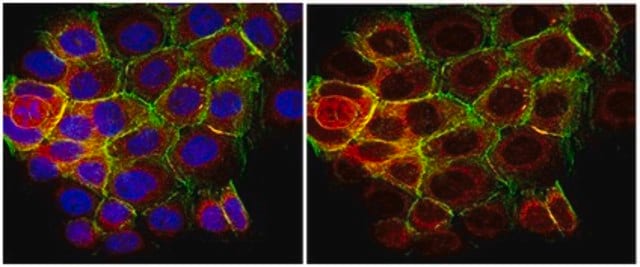
Immunofluorescence Analysis: A representative lot detected MLKL in Immunofluorescence applications (Samson, A.L., et al. (2021). Cell Death Differ. 28(7):2126-2144).
Western Blotting Analysis: A representative lot detected MLKL in Western Blotting applications (Samson, A.L., et al. (2021). Cell Death Differ. 28(7):2126-2144).
Note: Actual optimal working dilutions must be determined by end user as specimens, and experimental conditions may vary with the end user
Evaluated by Western Blotting in lysates from Mouse dermal fibroblasts.
Western Blotting Analysis (WB): A 1:500 dilution of this antibody detected MLKL in lysates from wild-type Mouse dermal fibroblasts, but not in lysates from fibroblasts with Mlkl knockout.
Tested Applications
Immunofluorescence Analysis: A representative lot detected MLKL in Immunofluorescence applications (Samson, A.L., et al. (2021). Cell Death Differ. 28(7):2126-2144).
Western Blotting Analysis: A representative lot detected MLKL in Western Blotting applications (Samson, A.L., et al. (2021). Cell Death Differ. 28(7):2126-2144).
Note: Actual optimal working dilutions must be determined by end user as specimens, and experimental conditions may vary with the end user
Anti-MLKL, clone 5A6, Cat. No. MABC1634, is a rat monoclonal antibody that detects MLKL and is tested for use in Immunofluorescence and Western Blotting.
物理的形状
Purified rat monoclonal antibody IgG2a in buffer containing 0.1 M Tris-Glycine (pH 7.4), 150 mM NaCl with 0.05% sodium azide
保管および安定性
Recommended storage: +2°C to +8°C.
その他情報
Concentration: Please refer to the Certificate of Analysis for the lot-specific concentration.
免責事項
Unless otherwise stated in our catalog or other company documentation accompanying the product(s), our products are intended for research use only and are not to be used for any other purpose, which includes but is not limited to, unauthorized commercial uses, in vitro diagnostic uses, ex vivo or in vivo therapeutic uses or any type of consumption or application to humans or animals.
適切な製品が見つかりませんか。
製品選択ツール.をお試しください
保管分類コード
12 - Non Combustible Liquids
WGK
WGK 1
引火点(°F)
Not applicable
引火点(℃)
Not applicable
適用法令
試験研究用途を考慮した関連法令を主に挙げております。化学物質以外については、一部の情報のみ提供しています。 製品を安全かつ合法的に使用することは、使用者の義務です。最新情報により修正される場合があります。WEBの反映には時間を要することがあるため、適宜SDSをご参照ください。
Jan Code
MABC1634-25UL:
MABC1634-100UL:
試験成績書(COA)
製品のロット番号・バッチ番号を入力して、試験成績書(COA) を検索できます。ロット番号・バッチ番号は、製品ラベルに「Lot」または「Batch」に続いて記載されています。
アクティブなフィルタ
ライフサイエンス、有機合成、材料科学、クロマトグラフィー、分析など、あらゆる分野の研究に経験のあるメンバーがおります。.
製品に関するお問い合わせはこちら(テクニカルサービス)