
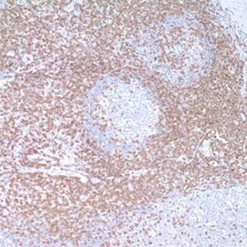


Based on the information available, this version does recognize the epsilon subunit. This conclusion is drawn from the fact that the CD3 epsilon, also known as the cytoplasmic CD3 antibody, exhibits a cytoplasmic staining pattern with perinuclear Golgi accentuation and very rarely membranous. Typically, this antibody is utilized for identifying T cell and NK cell lymphomas in FFPE tissues (please refer to representative staining photos). While the Cd3 surface antibody is commonly used in flow cytometry for membranous CD3 detection, the CD3 epsilon cytoplasmic is generally used in FFPE tissue, as is often done for IHC.
103R-9
CD3 (MRQ-39) Rabbit Monoclonal Antibody
Zaloguj sięWyświetlanie cen organizacyjnych i kontraktowych
About This Item
Kod UNSPSC:
12352203
NACRES:
NA.41
Produkt 103R-9 nie jest obecnie dostępny w sprzedaży w Twoim kraju Skontaktuj się z zespołem ds. pomocy technicznej
Polecane produkty
pochodzenie biologiczne
rabbit
Poziom jakości
100
500
białko sprzężone
unconjugated
forma przeciwciała
culture supernatant
rodzaj przeciwciała
primary antibodies
klon
MRQ-39, monoclonal
opis
For In Vitro Diagnostic Use in Select Regions (See Chart)
Formularz
buffered aqueous solution
reaktywność gatunkowa
human
opakowanie
pkg of 0.1 mL concentrate (103r-94)
pkg of 0.5 mL concentrate (103R-95)
pkg of 1.0 mL concentrate (103R-96)
pkg of 1.0 mL predilute (103R-97)
pkg of 7.0 mL predilute (103R-98)
producent / nazwa handlowa
Cell Marque®
IVD
for in vitro diagnostic use
metody
immunohistochemistry (formalin-fixed, paraffin-embedded sections): 1:100-1:500 (concentrated)
izotyp
IgG1
kontrola
tonsil
Warunki transportu
wet ice
temp. przechowywania
2-8°C
wizualizacja
membranous
informacje o genach
human ... CD3E(916)
Powiązane kategorie
Opis ogólny
Anti-CD3 has been considered the best all around T-cell marker. This antibody reacts with an antigen present in early thymocytes. The positive staining of this marker may represent a sign of early commitment to the T-Cell lineage.
Jakość
 IVD |  IVD |  IVD |  RUO |
Powiązanie
CD3 Positive Control Slides, Product No. 103S, are available for immunohistochemistry (formalin-fixed, paraffin-embedded sections).
Postać fizyczna
Solution in Tris Buffer, pH 7.3-7.7, with 1% BSA and <0.1% Sodium Azide
Uwaga dotycząca przygotowania
Download the IFU specific to your product lot and formatNote: This requires a keycode which can be found on your packaging or product label.
Inne uwagi
For Technical Service please contact: 800-665-7284 or email: [email protected]
Informacje prawne
Cell Marque is a registered trademark of Merck KGaA, Darmstadt, Germany
Ta strona może zawierać tekst przetłumaczony maszynowo.
Nie możesz znaleźć właściwego produktu?
Wypróbuj nasz Narzędzie selektora produktów.
Wybierz jedną z najnowszych wersji:
Certyfikaty analizy (CoA)
Lot/Batch Number
Nie widzisz odpowiedniej wersji?
Jeśli potrzebujesz konkretnej wersji, możesz wyszukać konkretny certyfikat według numeru partii lub serii.
Masz już ten produkt?
Dokumenty związane z niedawno zakupionymi produktami zostały zamieszczone w Bibliotece dokumentów.
E A Clark et al.
Immunology today, 10(7), 225-228 (1989-07-01)
During 1987, striking advances were made in defining the receptors and ligands for cell-to-cell adhesion interactions involving leukocytes. In 1988, two major leukocyte differentiation antigens, CD10 (cALLA) and CD45 (LCA, T200), were shown to be enzymes while two other markers
SM Denning, et al.
Leucocyte Typing III, 144-147 (1987)
Kennosuke Karube et al.
The American journal of surgical pathology, 27(10), 1366-1374 (2003-09-26)
We studied the morphologic, immunohistochemical, and clinical characteristics of 158 cases of lymphoblastic lymphoma. Based on immunophenotyping and cell lineage, cases were classified into B-cell type (CD20,CD19 or CD79a+, n = 53), T-cell type (surface CD3+, n = 84), and
P C Beverley et al.
European journal of immunology, 11(4), 329-334 (1981-04-01)
The properties of human lymphocyte fractions isolated either by sheep red cell(E) rosetting or by fluorescence-activated cell sorting after staining with UCHT1 monoclonal anti-T cell antibody have been compared. Two populations of E+ cells with very different phenotype and function
D Campana et al.
Journal of immunology (Baltimore, Md. : 1950), 138(2), 648-655 (1987-01-15)
Anti-CD3 (T3) Ab reacting with different proportions of thymocytes (anti-CD3a: UCHT1, anti-CD3b: T10B9, and anti-CD3c: OKT3) were tested for cytoplasmic (cCD3) and membrane (mCD3) expression in the bone marrow, thymus, and blood in man and selected primates. The expression of
-
Could you confirm if the CD3 clone (Catalog #103R-96) recognizes epsilon?
1 answer-
Helpful?
-
Active Filters
Nasz zespół naukowców ma doświadczenie we wszystkich obszarach badań, w tym w naukach przyrodniczych, materiałoznawstwie, syntezie chemicznej, chromatografii, analityce i wielu innych dziedzinach.
Skontaktuj się z zespołem ds. pomocy technicznej