Przejdź do zawartości
Merck
Wszystkie zdjęcia(1)

Dokumenty

MABT26

Sigma-Aldrich

Anti-E-cadherin Antibody, clone DECMA-1

clone Decma-1, from rat

Synonim(y):

Anti-Anti-Arc-1, Anti-Anti-BCDS1, Anti-Anti-CD324, Anti-Anti-CDHE, Anti-Anti-ECAD, Anti-Anti-LCAM, Anti-Anti-UVO

Zaloguj sięWyświetlanie cen organizacyjnych i kontraktowych


About This Item

Kod UNSPSC:
12352203
eCl@ss:
32160702
NACRES:
NA.41

pochodzenie biologiczne

rat

Poziom jakości

forma przeciwciała

purified antibody

rodzaj przeciwciała

primary antibodies

klon

Decma-1, monoclonal

reaktywność gatunkowa

human, mouse

metody

flow cytometry: suitable
immunocytochemistry: suitable
immunohistochemistry: suitable
immunoprecipitation (IP): suitable
western blot: suitable

izotyp

IgG1κ

numer dostępu NCBI

numer dostępu UniProt

Warunki transportu

dry ice

docelowa modyfikacja potranslacyjna

unmodified

informacje o genach

human ... CDH1(999)
mouse ... Cdh1(12550)

Opis ogólny

Cadherins are a family of transmembrane glycoproteins involved in Ca2+-dependent cell to cell interactions. The members of the family are differentially expressed in various tissues and function in the maintenance of tissue integrity, morphogenesis, and migration. Cadherins are divided into type I and type II subgroups. Type I cadherins include epithelial cadherin (E-cadherin), neural cadherin (N-cadherin), placental cadherin (P-cadherin) and retinal cadherin (R-cadherin). Type II include kidney cadherin (K-cadherin) and osteoblast cadherin (OB-cadherin). One of the best characterized cadherins is E-cadherin (epithelial cadherin or cadherin-1). It consists of calcium-binding extracellular domains responsible for interactions with other E-cadherin molecules on neighboring cells and interaction with integrin alphaEbeta7. The cytoplasmic domain of E-cadherin is linked to the actin cytoskeleton through cytoplasmic catenin proteins, thus establishing a complex localized to adherens junctions. In carcinomas, E-cadherin is frequently downregulated, consistent with its function of an invasion suppressor in normal epithelia.

Immunogen

Mouse embryonal carcinoma cell line PCC4

Zastosowanie

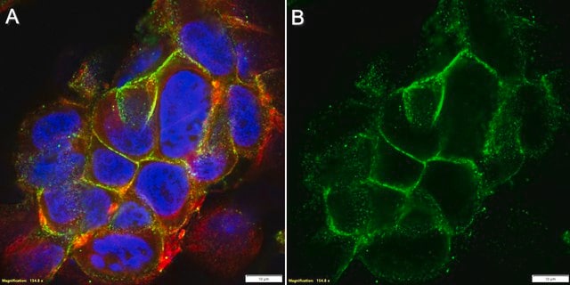
Immunohistochemistry Analysis: 1:300 dilution of this antibody has been shown to detect E-cadherin in mouse GI tract tissue.

Inhibition: A previous lot was used by an independent laboratory in inhibion studies, blocking E-cadherin associated cell adhesion. (Vestweber, 1985)

Flow Cytometry Analysis: A previous lot was used by an independent laboratory in flow cytometry applications. (Tang, 1994)
Research Category
Cell Structure
Research Sub Category
Adhesion (CAMs)
This Anti-E-cadherin Antibody, clone DECMA-1 is validated for use in WB, IH, IC, IP, FC, FUNC for the detection of E-cadherin.

Jakość

Evaluated by Western Blot in A431 cell lysate.

Western Blot Analysis: 1 µg/mL of this antibody detected E-Cadherin in 10 µg of A431 cell lysate.

Opis wartości docelowych

~ 106 kDa observed MW.

Postać fizyczna

Format: Purified
Protein G
Purified rat monoclonal IgG1κ in buffer containing PBS without preservative.

Przechowywanie i stabilność

Stable for 1 year at -20°C from date of receipt.
Handling Recommendations: Upon receipt and prior to removing the cap, centrifuge the vial and gently mix the solution. Aliquot into microcentrifuge tubes and store at -20°C. Avoid repeated freeze/thaw cycles, which may damage IgG and affect product performance.

Komentarz do analizy

Control
A431 cell lysate

Inne uwagi

Concentration: Please refer to the Certificate of Analysis for the lot-specific concentration.

Oświadczenie o zrzeczeniu się odpowiedzialności

Unless otherwise stated in our catalog or other company documentation accompanying the product(s), our products are intended for research use only and are not to be used for any other purpose, which includes but is not limited to, unauthorized commercial uses, in vitro diagnostic uses, ex vivo or in vivo therapeutic uses or any type of consumption or application to humans or animals.
This page may contain text that has been machine translated.

Not finding the right product?  

Try our Narzędzie selektora produktów.

Kod klasy składowania

12 - Non Combustible Liquids

Klasa zagrożenia wodnego (WGK)

WGK 2

Temperatura zapłonu (°F)

Not applicable

Temperatura zapłonu (°C)

Not applicable


Certyfikaty analizy (CoA)

Poszukaj Certyfikaty analizy (CoA), wpisując numer partii/serii produktów. Numery serii i partii można znaleźć na etykiecie produktu po słowach „seria” lub „partia”.

Masz już ten produkt?

Dokumenty związane z niedawno zakupionymi produktami zostały zamieszczone w Bibliotece dokumentów.

Odwiedź Bibliotekę dokumentów

S Pece et al.
The Journal of biological chemistry, 275(52), 41227-41233 (2000-09-02)
E-cadherins are well characterized cell surface molecules expressed in epithelial cells, which play a major role in cell adhesion through the establishment of calcium-dependent homophilic interactions at sites of cell-cell contacts. They are also integral components of morphogenetic programs controlling
Nicholas F Dybdal-Hargreaves et al.
Oncotarget, 10(45), 4719-4720 (2019-08-07)
[This corrects the article DOI: 10.18632/oncotarget.23798.].
Philippe Pujuguet et al.
Molecular biology of the cell, 14(5), 2181-2191 (2003-06-13)
Ezrin, a membrane cytoskeleton linker, is involved in cellular functions, including epithelial cell morphogenesis and adhesion. A mutant form of ezrin, ezrin T567D, maintains the protein in an open conformation, which when expressed in Madin-Darby canine kidney cells causes extensive
Zhibo Gai et al.
Journal of the American Society of Nephrology : JASN, 21(9), 1468-1476 (2010-05-29)
Mutations in TRPS1 cause tricho-rhino-pharyngeal syndrome (TRPS). Trps1 is essential for nephron development, acting downstream of Bmp7. Because Bmp7 counteracts epithelial-to-mesenchymal transition (EMT) and reverses chronic renal injury, we examined the function of Trps1 in renal fibrosis. Immunohistochemistry revealed Trps1
Marianela G Dalghi et al.
JCI insight, 6(19) (2021-09-01)
The mechanisms that link visceral mechanosensation to the perception of internal organ status (i.e., interoception) remain elusive. In response to bladder filling, the urothelium releases ATP, which is hypothesized to stimulate voiding function by communicating the degree of bladder fullness

Nasz zespół naukowców ma doświadczenie we wszystkich obszarach badań, w tym w naukach przyrodniczych, materiałoznawstwie, syntezie chemicznej, chromatografii, analityce i wielu innych dziedzinach.

Skontaktuj się z zespołem ds. pomocy technicznej