
価格・在庫情報は確認できません。詳細については、Merckをご覧ください。
おすすめの製品


由来生物
mouse
品質水準
抗体製品の状態
purified antibody
抗体製品タイプ
primary antibodies
クローン
1-146, monoclonal
交差性
human, rat
交差性(ホモロジーによる予測)
mouse
テクニック
immunohistochemistry: suitable
immunoprecipitation (IP): suitable
western blot: suitable
アイソタイプ
IgG1κ
NCBIアクセッション番号
UniProtアクセッション番号
輸送温度
wet ice
ターゲットの翻訳後修飾
unmodified
遺伝子情報
human ... STX3(6809)
rat ... Stx3(81802)
詳細
Syntaxin-3, also known as Stx3, is a member of the SNARE family which are complexes involved in membrane fusion. Syntaxin-3 is expressed in epithelial cells and is found apically in MDCK and Caco-2 cells (1).
特異性
This antibody recognizes Syntaxin-3.
免疫原
GST-tagged recombinant protein corresponding to residues contained within rat Syntaxin-3.
アプリケーション
Research Category
ニューロサイエンス
ニューロサイエンス
Research Sub Category
シナプス及びシナプス生物学
シナプス及びシナプス生物学
Detect Syntaxin-3 using this Anti-Syntaxin-3 Antibody, clone 1-146 validated for use in WB, IP, IH.
品質
Evaluated by Western Blot in rat brain tissue lysate.
Western Blot Analysis: : 0.5 µg/mL dilution of this antibody detected Syntaxin-3 on 15 µg of rat brain tissue lysate.
Western Blot Analysis: : 0.5 µg/mL dilution of this antibody detected Syntaxin-3 on 15 µg of rat brain tissue lysate.
ターゲットの説明
Approx. 33 kDa
物理的形状
Protein G Purified
Format: Purified
Purified mouse monoclonal in buffer containing 0.1 M Tris-glycine, pH 7.4, 150 mM NaCl with 0.05% NaN3.
保管および安定性
Maintain refrigerated at 2-8°C for 1 year from date of receipt.
アナリシスノート
Control
Rat brain tissue lysate
Rat brain tissue lysate
その他情報
Concentration: Please refer to the Certificate of Analysis for the lot-specific concentration.
免責事項
Unless otherwise stated in our catalog or other company documentation accompanying the product(s), our products are intended for research use only and are not to be used for any other purpose, which includes but is not limited to, unauthorized commercial uses, in vitro diagnostic uses, ex vivo or in vivo therapeutic uses or any type of consumption or application to humans or animals.
適切な製品が見つかりませんか。
製品選択ツール.をお試しください
保管分類コード
12 - Non Combustible Liquids
WGK
WGK 1
引火点(°F)
Not applicable
引火点(℃)
Not applicable
適用法令
試験研究用途を考慮した関連法令を主に挙げております。化学物質以外については、一部の情報のみ提供しています。 製品を安全かつ合法的に使用することは、使用者の義務です。最新情報により修正される場合があります。WEBの反映には時間を要することがあるため、適宜SDSをご参照ください。
Jan Code
MAB2258:
試験成績書(COA)
製品のロット番号・バッチ番号を入力して、試験成績書(COA) を検索できます。ロット番号・バッチ番号は、製品ラベルに「Lot」または「Batch」に続いて記載されています。
Adrian J Giovannone et al.
The Journal of biological chemistry, 293(15), 5478-5491 (2018-02-25)
Syntaxins are a conserved family of SNARE proteins and contain C-terminal transmembrane anchors required for their membrane fusion activity. Here we show that Stx3 (syntaxin 3) unexpectedly also functions as a nuclear regulator of gene expression. We found that alternative
Daniëlle Rianne José Verboogen et al.
eLife, 6 (2017-05-20)
SNARE proteins play a crucial role in intracellular trafficking by catalyzing membrane fusion, but assigning SNAREs to specific intracellular transport routes is challenging with current techniques. We developed a novel Förster resonance energy transfer-fluorescence lifetime imaging microscopy (FRET-FLIM)-based technique allowing
Ritika Chatterjee et al.
Traffic (Copenhagen, Denmark), 24(7), 270-283 (2023-04-28)
Intracellular membrane fusion is mediated by membrane-bridging complexes of soluble N-ethylmaleimide-sensitive factor attachment protein receptors (SNAREs). SNARE proteins are one of the key players in vesicular transport. Several reports shed light on intracellular bacteria modulating host SNARE machinery to establish
Ying Hsu et al.
Molecular therapy. Nucleic acids, 31, 164-181 (2023-01-27)
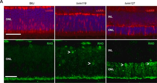
Blindness in Bardet-Biedl syndrome (BBS) is caused by dysfunction and loss of photoreceptor cells in the retina. BBS10, mutations of which account for approximately 21% of all BBS cases, encodes a chaperonin protein indispensable for the assembly of the BBSome
Hiroshi Gomi et al.
The journal of histochemistry and cytochemistry : official journal of the Histochemistry Society, 65(11), 637-653 (2017-09-16)
The comparative structure and expression of salivary components and vesicular transport proteins in the canine major salivary glands were investigated. Histochemical analysis revealed that the morphology of the five major salivary glands-parotid, submandibular, polystomatic sublingual, monostomatic sublingual, and zygomatic glands-was
アクティブなフィルタ
ライフサイエンス、有機合成、材料科学、クロマトグラフィー、分析など、あらゆる分野の研究に経験のあるメンバーがおります。.
製品に関するお問い合わせはこちら(テクニカルサービス)