MABT366
Anti-Claudin-1/CLDN1 Antibody, clone 7A5
clone 7A5, from mouse
Szinonimák:
Claudin-1, Senescence-associated epithelial membrane protein
About This Item
ICC
WB
immunocytochemistry: suitable
western blot: suitable
Javasolt termékek
biológiai forrás
mouse
Minőségi szint
antitest forma
purified immunoglobulin
antitest terméktípus
primary antibodies
klón
7A5, monoclonal
faj reaktivitás
human
nem léphet reakcióba
mouse
technika/technikák
flow cytometry: suitable
immunocytochemistry: suitable
western blot: suitable
izotípus
IgG1κ
NCBI elérési szám
UniProt elérési szám
kiszállítva
dry ice
célzott transzláció utáni módosítás
unmodified
Géninformáció
human ... CLDN1(9076) , NISCH(11188)
Általános leírás
Egyediség
Immunogén
Alkalmazás
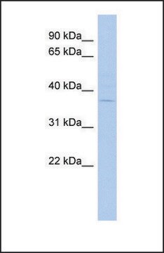
Western Blotting Analysis: 0.5 µg/mL from a representative lot detected exogenously expressed claudin-1 in CLDN1-transfected, but not mock-transfected, HT1080 cells (Courtesy of Professor Masuo Kondoh, PhD, Osaka University, Japan).
Immunocytochemistry Analysis: A representative lot immunostained the surface of Huh-7.5.1 human hepatoma cells, but not the non-claudin-1-/CLDN1-expressing S7-A cells (Fukasawa, M., et al. (2015). J. Virol. 89(9):4866-4879).
Flow Cytometry Analysis: A representative lot specifically immunostained HT1080 cells expressing exogenously transfected human claudin-1 (CLDN1), but not HT1080 cells expressing human claudin-2, -4, -5, -6, -7, or -9, nor L cells expressing mouse claudin-1 (Fukasawa, M., et al. (2015). J. Virol. 89(9):4866-4879).
Flow Cytometry Analysis: A representative lot immunostained HEK293T transfectants expressing FLAG-tagged human claudin-1/CLDN1 and human-mouse claudin-1 chimeras with the second human extracellular loop (EL2), but not chimeras with the mouse EL2. M152L, but not V155I, mutation in human EL2 abolished the immunoreactivity (Fukasawa, M., et al. (2015). J. Virol. 89(9):4866-4879).
Western Blotting Analysis: A representative lot detected FLAG-tagged human claudin-1/CLDN1 and human-mouse claudin-1 chimeras with the second human extracellular loop (EL2), but not chimeras with the mouse EL2. M152L, but not V155I, mutation in the second human extracellular loop abolished the immunoreactivity (Fukasawa, M., et al. (2015). J. Virol. 89(9):4866-4879).
ELISA Analysis: A representative lot detected claudin-1/CLDN1 immunoreactivity in 3.7% formaldehyde-fixed Huh-7.5.1 human hepatoma cells by "cell ELISA" (Fukasawa, M., et al. (2015). J. Virol. 89(9):4866-4879).
Neutralization Analysis: A representative lot inhibited HCV infection of cultured Huh-7.5.1 human hepatoma cells in vitro and of human liver-chimeric mice in vivo (Fukasawa, M., et al. (2015). J. Virol. 89(9):4866-4879).
Cell Structure
Infectious Diseases - Viral
Minőség
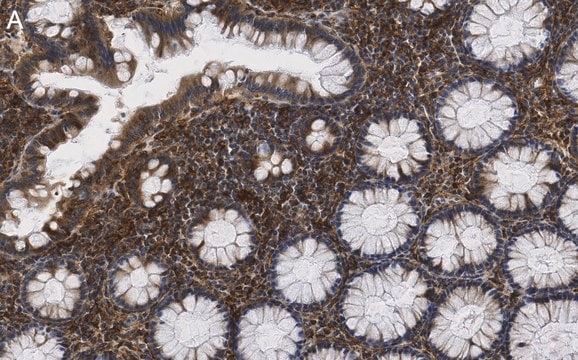
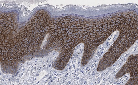
Immunocytochemistry Analysis: 10 µg/mL of this antibody detected Claudin-1/CLDN1 in HepG2 cells.
Cél megnevezése
Fizikai forma
Tárolás és stabilitás
Handling Recommendations: Upon receipt and prior to removing the cap, centrifuge the vial and gently mix the solution. Aliquot into microcentrifuge tubes and store at -20°C. Avoid repeated freeze/thaw cycles, which may damage IgG and affect product performance.
Egyéb megjegyzések
Jogi nyilatkozat
Nem találja a megfelelő terméket?
Próbálja ki a Termékválasztó eszköz. eszközt
javasolt
Tárolási osztály kódja
12 - Non Combustible Liquids
WGK
WGK 2
Lobbanási pont (F)
Not applicable
Lobbanási pont (C)
Not applicable
Analitikai tanúsítványok (COA)
Analitikai tanúsítványok (COA) keresése a termék sarzs-/tételszámának megadásával. A sarzs- és tételszámok a termék címkéjén találhatók, a „Lot” vagy „Batch” szavak után.
Már rendelkezik ezzel a termékkel?
Az Ön által nemrégiben megvásárolt termékekre vonatkozó dokumentumokat a Dokumentumtárban találja.
Tudóscsoportunk valamennyi kutatási területen rendelkezik tapasztalattal, beleértve az élettudományt, az anyagtudományt, a kémiai szintézist, a kromatográfiát, az analitikát és még sok más területet.
Lépjen kapcsolatba a szaktanácsadással

