Ugrás a tartalomra
Merck
Összes fotó(1)

Fontos dokumentumok

MABC1595

Sigma-Aldrich

Anti-RIPK3/RIP3 Antibody, clone 8G7

Szinonimák:

Anti-RIPK3 Antibody, Clone 8G7 Anti-RIPK3, RIPK3 Detection Antibody

Bejelentkezésa Szervezeti és Szerződéses árazás megtekintéséhez


About This Item

UNSPSC kód:
12352203
NACRES:
NA.43
konjugátum:
unconjugated
application:
IF
WB
klón:
8G7, monoclonal
faj reaktivitás:
mouse
citations:
technika/technikák:
immunofluorescence: suitable
western blot: suitable

biológiai forrás

rat

Minőségi szint

konjugátum

unconjugated

antitest forma

purified antibody

antitest terméktípus

primary antibodies

klón

8G7, monoclonal

molekulatömeg

calculated mol wt 53.32 kDa
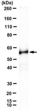
observed mol wt ~53 kDa

faj reaktivitás

mouse

kiszerelés

antibody small pack of 100 μL

technika/technikák

immunofluorescence: suitable
western blot: suitable

izotípus

IgG2aκ

UniProt elérési szám

kiszállítva

dry ice

tárolási hőmérséklet

2-8°C

célzott transzláció utáni módosítás

unmodified

Általános leírás

Receptor-interacting serine/threonine-protein kinase 3 (UniProt: EC:2.7.11.1, RIP-like protein kinase 3, Receptor-interacting protein 3, RIP-3, mRIP3) is encoded by the Ripk3 (also known as Rip3) gene (Gene ID: 56532) in murine species. RIPK3 is a serine/threonine protein kinase that is expressed in embryo and adult spleen, liver, testis, brain, and lung. Its protein kinase domain is localized in amino acids 22 to 293 in the N-terminal half and it also has a C-terminal RIP homolytic interaction motif (RHIM; aa 440-461), which mediates interaction with the RHIM motif of RIPK1 and facilitate the formation of a necrosome. RIPK3 is shown to activate both necroptosis and apoptosis. Activated RIPK3 forms a necrosis-inducing complex and mediates phosphorylation of MLKL, promoting MLKL localization to the plasma membrane and execution of programmed necrosis that is characterized by calcium influx and plasma membrane damage. Its location is mainly cytoplasmic, however, following infection, it can also be detected in the nucleus. RIPK3 also regulates apoptosis that is dependent on RIPK1, FADD, and CASP8, and is independent of MLKL and RIPK3 kinase activity. It phosphorylates RIPK1 and then RIPK1 and RIPK3 undergo reciprocal auto- and trans-phosphorylation. In some cell types, RIPK3 is also reported to restrict viral replication by promoting cell death-independent responses. Clone 8G7 is raised against an extended kinase domain (residues 2-353) of mouse RIPK3 and can detect forms that may not be detected by antibodies directed towards the C-terminus of the full-length RIPK3. (Ref.: Samson, AL., et al. (2021). Cell Death Differ. https://doi.org/10.1038/s41418-021-00742-x; Petrie, EJ., et al. (2019). Cell Rep. 28(13); 3309-3319).

Egyediség

Clone 8G7 is a rat monoclonal antibody that detects murine Receptor-interacting serine/threonine-protein kinase 3 (RIPK3). It targets an epitope within the extended kinase domain that includes most of the N-terminal half and a part of C-terminal half.

Immunogén

Recombinant fragment corresponding to 352 amino acids from the extended kinase domain of murine Receptor-interacting serine/threonine-protein kinase 3 (RIPK3).

Alkalmazás

Quality Control Testing

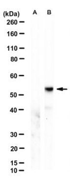
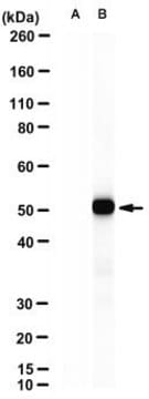
Evaluated by Western Blotting in Mouse dermal fibroblast lysates.

Western Blotting Analysis: A 1:500 dilution of this antibody detected RIPK3/RIP3 in Mouse dermal fibroblast lysates.

Tested Applications

Western Blotting Analysis: A representative lot detected RIPK3/RIP3 in Western Blotting applications (Petrie, E.J., et. al. (2019). Cell Rep. 28(13):3309-3319).

Immunofluorescence Analysis: A representative lot detected RIPK3/RIP3 in Immunofluorescence applications (Samson, A.L., et. al. (2021). Cell Death Differ. doi: 10.1038/s41418-021-00742-x)

Note: Actual optimal working dilutions must be determined by end user as specimens, and experimental conditions may vary with the end user
Anti-RIPK3/RIP3, clone 8G7, Cat. No. MABC1595, is a rat monoclonal antibody that detects RIPK3/RIP3 and is tested for use in Immunofluorescence and Western Blotting.

Fizikai forma

Purified rat monoclonal antibody IgG2a in buffer containing 0.1 M Tris-Glycine (pH 7.4), 150 mM NaCl with 0.05% sodium azide.

Tárolás és stabilitás

Recommend storage at +2°C to +8°C. For long term storage antibodies can be kept at -20°C. Avoid repeated freeze-thaws.

Egyéb megjegyzések

Concentration: Please refer to the Certificate of Analysis for the lot-specific concentration.

Jogi nyilatkozat

Unless otherwise stated in our catalog or other company documentation accompanying the product(s), our products are intended for research use only and are not to be used for any other purpose, which includes but is not limited to, unauthorized commercial uses, in vitro diagnostic uses, ex vivo or in vivo therapeutic uses or any type of consumption or application to humans or animals.

Nem találja a megfelelő terméket?  

Próbálja ki a Termékválasztó eszköz. eszközt

Tárolási osztály kódja

12 - Non Combustible Liquids

WGK

WGK 1

Lobbanási pont (F)

Not applicable

Lobbanási pont (C)

Not applicable


Analitikai tanúsítványok (COA)

Analitikai tanúsítványok (COA) keresése a termék sarzs-/tételszámának megadásával. A sarzs- és tételszámok a termék címkéjén találhatók, a „Lot” vagy „Batch” szavak után.

Már rendelkezik ezzel a termékkel?

Az Ön által nemrégiben megvásárolt termékekre vonatkozó dokumentumokat a Dokumentumtárban találja.

Dokumentumtár megtekintése

Tudóscsoportunk valamennyi kutatási területen rendelkezik tapasztalattal, beleértve az élettudományt, az anyagtudományt, a kémiai szintézist, a kromatográfiát, az analitikát és még sok más területet.

Lépjen kapcsolatba a szaktanácsadással