
HPA024223
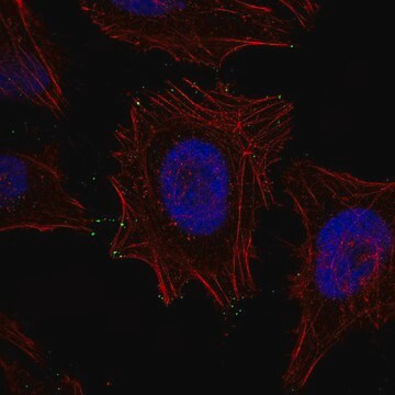
Anti-MYO10 antibody produced in rabbit
Prestige Antibodies® Powered by Atlas Antibodies, affinity isolated antibody, buffered aqueous glycerol solution
Sinônimo(s):
Anti-Myosin-X, Anti-Unconventional myosin-10
Selecione um tamanho
R$ 4.140,00
Selecione um tamanho
About This Item
R$ 4.140,00
Produtos recomendados


fonte biológica
rabbit
Nível de qualidade
conjugado
unconjugated
forma do anticorpo
affinity isolated antibody
tipo de produto de anticorpo
primary antibodies
clone
polyclonal
linha de produto
Prestige Antibodies® Powered by Atlas Antibodies
Formulário
buffered aqueous glycerol solution
reatividade de espécies
mouse, human
técnica(s)
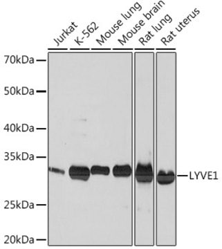
immunofluorescence: 0.25-2 μg/mL
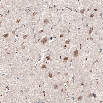
immunohistochemistry: 1:500-1:1000
sequência de imunogênio
QRMKEQQELSLTEASLQKLQERRDQELRRLEEEACRAAQEFLESLNFDEIDECVRNIERSLSVGSEFSSELAESACEEKPNFNFSQPYPEEEVDEGFEADDDAFKDSPNPSEHGHSDQRTSGIRTSDDSSEEDPYMNDTVVPTSPSA
nº de adesão UniProt
Condições de expedição
wet ice
temperatura de armazenamento
−20°C
modificação pós-traducional do alvo
unmodified
Informações sobre genes
human ... MYO10(4651)
Descrição geral
Imunogênio
Aplicação
Anti-MYO10 antibody produced in rabbit has ben used in western blotting and immunofluorescence.[1][2]
Ações bioquímicas/fisiológicas
Características e benefícios
Every Prestige Antibody is tested in the following ways:
- IHC tissue array of 44 normal human tissues and 20 of the most common cancer type tissues.
- Protein array of 364 human recombinant protein fragments.
Ligação
forma física
Informações legais
Exoneração de responsabilidade
Não está encontrando o produto certo?
Experimente o nosso Ferramenta de seleção de produtos.
Código de classe de armazenamento
10 - Combustible liquids
Classe de risco de água (WGK)
WGK 1
Ponto de fulgor (°F)
Not applicable
Ponto de fulgor (°C)
Not applicable
Escolha uma das versões mais recentes:
Já possui este produto?
Encontre a documentação dos produtos que você adquiriu recentemente na biblioteca de documentos.
Active Filters
Nossa equipe de cientistas tem experiência em todas as áreas de pesquisa, incluindo Life Sciences, ciência de materiais, síntese química, cromatografia, química analítica e muitas outras.
Entre em contato com a assistência técnica