HPA041163
Anti-ENKD1 antibody produced in rabbit

Prestige Antibodies® Powered by Atlas Antibodies, affinity isolated antibody, buffered aqueous glycerol solution
Synonim(y):
Anti-C16orf48, Anti-Chromosome 16 open reading frame 48, Anti-Dkfzp434a1319
About This Item
Polecane produkty
pochodzenie biologiczne
rabbit
Poziom jakości
białko sprzężone
unconjugated
forma przeciwciała
affinity isolated antibody
rodzaj przeciwciała
primary antibodies
klon
polyclonal
linia produktu
Prestige Antibodies® Powered by Atlas Antibodies
Formularz
buffered aqueous glycerol solution
reaktywność gatunkowa
human
rozszerzona walidacja
recombinant expression
Learn more about Antibody Enhanced Validation
metody
immunoblotting: 0.04-0.4 μg/mL
immunofluorescence: 0.25-2 μg/mL
immunohistochemistry: 1:200-1:500
sekwencja immunogenna
EGNALKLDLLTSDRALDTTAPRGPCIGPGAGEILERGQRGVGDVLLQLEGISLGPGASLKRKDPKDHEKENL
Warunki transportu
wet ice
temp. przechowywania
−20°C
docelowa modyfikacja potranslacyjna
unmodified
informacje o genach
human ... C16orf48(84080)
Immunogen
Zastosowanie
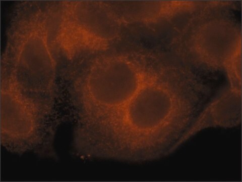
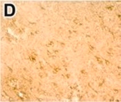
The Human Protein Atlas project can be subdivided into three efforts: Human Tissue Atlas, Cancer Atlas, and Human Cell Atlas. The antibodies that have been generated in support of the Tissue and Cancer Atlas projects have been tested by immunohistochemistry against hundreds of normal and disease tissues and through the recent efforts of the Human Cell Atlas project, many have been characterized by immunofluorescence to map the human proteome not only at the tissue level but now at the subcellular level. These images and the collection of this vast data set can be viewed on the Human Protein Atlas (HPA) site by clicking on the Image Gallery link. We also provide Prestige Antibodies® protocols and other useful information.
Cechy i korzyści
Every Prestige Antibody is tested in the following ways:
- IHC tissue array of 44 normal human tissues and 20 of the most common cancer type tissues.
- Protein array of 364 human recombinant protein fragments.
Powiązanie
Postać fizyczna
Informacje prawne
Nie możesz znaleźć właściwego produktu?
Wypróbuj nasz Narzędzie selektora produktów.
Kod klasy składowania
10 - Combustible liquids
Klasa zagrożenia wodnego (WGK)
WGK 1
Temperatura zapłonu (°F)
Not applicable
Temperatura zapłonu (°C)
Not applicable
Wybierz jedną z najnowszych wersji:
Certyfikaty analizy (CoA)
Nie widzisz odpowiedniej wersji?
Jeśli potrzebujesz konkretnej wersji, możesz wyszukać konkretny certyfikat według numeru partii lub serii.
Masz już ten produkt?
Dokumenty związane z niedawno zakupionymi produktami zostały zamieszczone w Bibliotece dokumentów.
Nasz zespół naukowców ma doświadczenie we wszystkich obszarach badań, w tym w naukach przyrodniczych, materiałoznawstwie, syntezie chemicznej, chromatografii, analityce i wielu innych dziedzinach.
Skontaktuj się z zespołem ds. pomocy technicznej


