
Przejdź do
A1226
Anti-Actopaxin antibody produced in rabbit
IgG fraction of antiserum, PBS solution
Synonim(y):
Anti-α-Parvin, Anti-CH-ILKBP
Informacje o tej pozycji
biological source
rabbit
Quality Level
conjugate
unconjugated
antibody form
IgG fraction of antiserum
antibody product type
primary antibodies
clone
polyclonal
form
PBS solution
mol wt
antigen 42 kDa
species reactivity
human, mouse
technique(s)
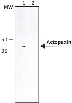
microarray: suitable, western blot: 1:3,000 using whole cell extract of the human endothelial ECV304 cell line.
UniProt accession no.
shipped in
dry ice
storage temp.
−20°C
target post-translational modification
unmodified
Gene Information
human ... PARVA(55742)
mouse ... Parva(57342)
General description
Rabbit anti-actopaxin recognizes actopaxin (42 kDa). Staining of actopaxin in immunoblotting is specifically inhibited with actopaxin immunizing peptide (mouse, amino acids 35-53).
Immunogen
Application
Immunofluorescence (1 paper)
Physical form
Disclaimer
1 of 1
Ta pozycja | |||
|---|---|---|---|
| conjugate unconjugated | conjugate unconjugated | conjugate unconjugated | conjugate unconjugated |
| biological source rabbit | biological source rabbit | biological source rabbit | biological source rabbit |
| antibody form IgG fraction of antiserum | antibody form IgG fraction of antiserum | antibody form affinity isolated antibody | antibody form IgG fraction of antiserum |
| Quality Level 200 | Quality Level 200 | Quality Level 200 | Quality Level - |
| clone polyclonal | clone polyclonal | clone polyclonal | clone polyclonal |
| technique(s) microarray: suitable, western blot: 1:3,000 using whole cell extract of the human endothelial ECV304 cell line. | technique(s) western blot: 1:2,000-1:4,000 using whole extracts of HeLa human epithelioid carcinoma and mouse NIH3T3 cells | technique(s) immunocytochemistry: 1-2 μg/mL using cultured chicken fibroblasts, immunohistochemistry (formalin-fixed, paraffin-embedded sections): 2-4 μg/mL using sections of human appendix, mouse heart, and frog skeletal muscle, indirect immunofluorescence: suitable, western blot: 0.5-1 μg/mL using whole extract of the human epitheloid carcinoma HeLa cell line., western blot: 2-4 ng/mL using whole extract of rat skeletal muscle | technique(s) western blot: 1:500-1:1,000 using whole extracts of human SH-SY5Y cells, mouse brain, and rat brain |


Still not finding the right product?
Wypróbuj nasze narzędzie Narzędzie selektora produktów, aby zawęzić opcje.
Wybierz jedną z najnowszych wersji:
Masz już ten produkt?
Dokumenty związane z niedawno zakupionymi produktami zostały zamieszczone w Bibliotece dokumentów.
Powiązane treści
Datasheet
Numer pozycji handlu globalnego
| SKU | NUMER GTIN |
|---|---|
| A1226-200UL | 04061837871177 |