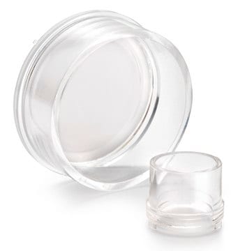
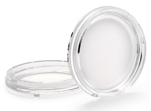
PICM01250
Millicell®スタンディング細胞培養インサート
pore size 0.4 μm, diam. 12 mm, transparent PTFE membrane, hydrophilic, size 24 wells, sterile
別名:
Millicell細胞培養インサート 12 mm 親水性PTFE 0.4 µm, 細胞培養インサート, 組織培養インサート, 組織培養プレート, 透過性培養インサート
About This Item
おすすめの製品
物質
polystyrene housing
transparent PTFE membrane
品質水準
無菌性
ethylene oxide treated
sterile
特徴
hydrophilic
包装
pack of 50
メーカー/製品名
Millicell®
パラメーター
50 °C max. temp.
テクニック
cell attachment: suitable
cell culture | mammalian: suitable
cell differentiation: suitable
H
10.5 mm
直径
12 mm
ろ過面積
0.6 cm2
サイズ
24 wells
表面積
0.6 cm2
有効容積
0.6 mL
色
transparent, when wetted
マトリックス
Biopore™
ポアサイズ
0.4 μm pore size
結合型
low binding surface
検出方法
fluorometric
輸送温度
ambient
関連するカテゴリー
詳細
Millicell®インサートにより、接着細胞または懸濁細胞が頂端側と側底側の両方から培地にアクセスできます。細胞の増殖、構造、機能は、in vivoで生じることをより厳密に模倣します。また、Millicell®インサートでは、細胞単層の両側を検討することが可能です。
Millicell®スタンディングインサート:
- 良好な細胞増殖を促進し、細胞研究のすばらしい機会が得られます
メンブレンタイプ:
Bioporeメンブレン(親水性PTFE)
- 低タンパク質結合、生細胞観察、および免疫蛍光染色アプリケーション用
アプリケーション
細胞接着、細胞増殖、細胞分化、免疫細胞染色
アプリケーション
細胞培養
包装
法的情報
保管分類コード
10-13 - German Storage Class 10 to 13
試験成績書(COA)
製品のロット番号・バッチ番号を入力して、試験成績書(COA) を検索できます。ロット番号・バッチ番号は、製品ラベルに「Lot」または「Batch」に続いて記載されています。
この製品を見ている人はこちらもチェック
資料
16HBE14o- human bronchial epithelial cells used to model respiratory epithelium for the research of cystic fibrosis, viral pulmonary pathology (SARS-CoV), asthma, COPD, effects of smoking and air pollution. See over 5k publications.
プロトコル
Toluidine blue selectively stains nuclear material and acidic tissue components, aiding in histological staining for tissues rich in DNA/RNA.
3D cell culture protocol for generating epidermal human skin tissue using primary human keratinocytes, dermal fibroblasts, and collagen-coated transwell inserts.
This protocol covers 3 modes for the microscopic examination of cell samples.
This page covers the basic indirect co-culture procedure on both sides of Millicell cell culture insert membranes.
関連コンテンツ
Streamline TEER data capture and analysis with user-friendly enhancements including an intuitive touchscreen interface, standing in-well probe, and automatic data logging.
ライフサイエンス、有機合成、材料科学、クロマトグラフィー、分析など、あらゆる分野の研究に経験のあるメンバーがおります。.
製品に関するお問い合わせはこちら(テクニカルサービス)


