Direkt zum Inhalt
Merck
Alle Fotos(1)

Wichtige Dokumente

MABN827

Sigma-Aldrich

Anti Tau (MAPT) Antibody (not human)

mouse monoclonal, T49

Synonym(e):

Microtubule-associated protein tau, Neurofibrillary tangle protein, Paired helical filament-tau, PHF-tau

Anmeldenzur Ansicht organisationsspezifischer und vertraglich vereinbarter Preise


About This Item

UNSPSC-Code:
12352203
eCl@ss:
32160702
NACRES:
NA.41
Preise und Verfügbarkeit sind derzeit nicht verfügbar.

Produktbezeichnung

Anti-Tau Antibody, clone T49 (Not human), clone T49, from mouse

Biologische Quelle

mouse

Qualitätsniveau

Antikörperform

purified antibody

Antikörper-Produkttyp

primary antibodies

Klon

T49, monoclonal

Speziesreaktivität

mouse, rat, bovine

Darf nicht reagieren mit

human

Methode(n)

immunohistochemistry: suitable
western blot: suitable

Isotyp

IgG1κ

NCBI-Hinterlegungsnummer

UniProt-Hinterlegungsnummer

Versandbedingung

wet ice

Posttranslationale Modifikation Target

unmodified

Angaben zum Gen

Allgemeine Beschreibung

Tau or Microtubule-associated protein tau (MAPT), also known as neurofibrillary tangle protein and paired helical filament-tau (PHF-tau), is a cytosolic protein that promotes microtubule assembly and stability, and might be involved in the establishment and maintenance of axonal polarity in neurons. Tau binds to and is thought to function as a linker protein between axonal microtubules and neural plasma membrane components. There are multiple isoforms, and the short isoforms allow plasticity of the cytoskeleton whereas the longer isoforms may preferentially play a role in its stabilization. PAD is the phosphatase activating domain, and has been demonstrated to be involved in the inhibition of anterograde, kinesin-based fast axonal transport (FAT) by activating axonal protein phosphatase 1 (PP1) and glycogen synthase kinase 3 (GSK3), independent of microtubule binding. Defects in Tau are thought to be the cause of a number of neurodegenerative diseases, including frontotemporal dementia (FTD), pallido-ponto-nigral degeneration (PPND), Pick disease of the brain (PIDB), corticobasal degeneration (CBD), supranuclear palsy type 1 (PSNP1), Alzheimer disease, and Parkinson disease. Clone T49 exhibits immunoreactivity against bovine, rat, and murine, but not human, Tau (UniProt P29172, P19332, P10637, P10636, respectively).

Immunogen

Purified corresponding to bovine Tau.

Anwendung

Anti-Tau Antibody, clone T49 (Not human) is an antibody against Tau for use in Western Blotting and Immunohistochemistry.
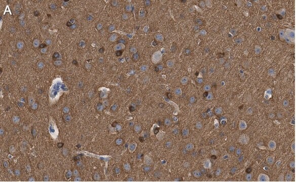
Immunohistochemistry Analysis: A 1:1,000 dilution from a representative lot detected Tau in mouse cerebral cortex, mouse kidney, and mouse small intestine tissue.
Western Blotting Analysis: A representative lot detected Tau in PS19 neurons treated with PBS or transduced with strain A or strain B FL a-syn pffs (Guo, J.L., et al. (2013). Cell. 154:103-117).

Qualität

Evaluated by Western Blotting in mouse and human brain tissue lysate.

Western Blotting Analysis: 0.5 µg/mL of this antibody detected Tau in 10 µg of mouse and human brain tissue lysate.

Zielbeschreibung

~53 kDa observed. This protein has multiple isoforms which range from 45-76 kDa

Physikalische Form

Format: Purified

Sonstige Hinweise

Concentration: Please refer to lot specific datasheet.

Sie haben nicht das passende Produkt gefunden?  

Probieren Sie unser Produkt-Auswahlhilfe. aus.

Lagerklassenschlüssel

12 - Non Combustible Liquids

WGK

WGK 1

Flammpunkt (°F)

Not applicable

Flammpunkt (°C)

Not applicable


Analysenzertifikate (COA)

Suchen Sie nach Analysenzertifikate (COA), indem Sie die Lot-/Chargennummer des Produkts eingeben. Lot- und Chargennummern sind auf dem Produktetikett hinter den Wörtern ‘Lot’ oder ‘Batch’ (Lot oder Charge) zu finden.

Besitzen Sie dieses Produkt bereits?

In der Dokumentenbibliothek finden Sie die Dokumentation zu den Produkten, die Sie kürzlich erworben haben.

Die Dokumentenbibliothek aufrufen

Sheng Chen et al.
Nature communications, 15(1), 2436-2436 (2024-03-19)
Parkinson's disease (PD) is closely linked to α-synuclein (α-syn) misfolding and accumulation in Lewy bodies. The PDZ serine protease HTRA1 degrades fibrillar tau, which is associated with Alzheimer's disease, and inactivating mutations to mitochondrial HTRA2 are implicated in PD. Here
Michael Fassler et al.
Cells, 12(11) (2023-06-10)
TREM2 is a membrane receptor expressed on microglia that plays a pivotal role in the organization and function of these innate immune cell components within the neurodegenerated brain. Whereas TREM2 deletion has been studied extensively in experimental beta-amyloid and Tau-based
Alberto Carpinteiro Soares et al.
The Journal of biological chemistry, 296, 100636-100636 (2021-04-09)
Tauopathies, such as Alzheimer's disease (AD), are neurodegenerative disorders characterized by the deposition of hyperphosphorylated tau aggregates. Proteopathic tau seeds spread through the brain in a temporospatial pattern, indicative of transsynaptic propagation. It is hypothesized that reducing the uptake of
Sarah Helena Nies et al.
The Journal of biological chemistry, 297(4), 101159-101159 (2021-09-05)
In Alzheimer's disease (AD), deposition of pathological tau and amyloid-β (Aβ) drive synaptic loss and cognitive decline. The injection of misfolded tau aggregates extracted from human AD brains drives templated spreading of tau pathology within WT mouse brain. Here, we
Parissa Fereydouni-Forouzandeh et al.
Methods in molecular biology (Clifton, N.J.), 2754, 309-321 (2024-03-21)
Tau is a microtubule-associated protein enriched in the axonal compartment. Its most well-known function is to bind and stabilize microtubules. In Alzheimer's disease and other neurodegenerative diseases known as tauopathies, tau undergoes several abnormal post-translational modifications including hyperphosphorylation, conformational changes

Questions

  1. Can nucleolar murine tau be detected using this antibody? Do you have an antibody that does detect nucleus-localized murine tau?

    1 answer
    1. Nuclear staining in mice has not been observed with this antibody. The immunohistochemistry images provided show cytoplasmic staining of glial cells, extracellular matrix staining of mouse cerebral cortex, membrane and cytoplasmic staining of tubule epithelial cells and podocytes in glomeruli of mouse kidney, and cytoplasmic staining of neurons in the muscular layer of mouse small intestine tissues. Mouse tau antibodies have not been validated for nuclear localization.

      Helpful?

Reviews

No rating value

Active Filters

Unser Team von Wissenschaftlern verfügt über Erfahrung in allen Forschungsbereichen einschließlich Life Science, Materialwissenschaften, chemischer Synthese, Chromatographie, Analytik und vielen mehr..

Setzen Sie sich mit dem technischen Dienst in Verbindung.