Direkt zum Inhalt
Merck
Alle Fotos(1)

Dokumente

MABF939

Sigma-Aldrich

Anti-IFNAR1 Antibody, clone 4G8

clone 4G8, from mouse

Synonym(e):

Interferon alpha/beta receptor 1, CRF2-1, Cytokine receptor class-II member 1, Cytokine receptor family 2 member 1, IFN-alpha/beta receptor 1, IFN-R-1, Type I interferon receptor 1

Anmeldenzur Ansicht organisationsspezifischer und vertraglich vereinbarter Preise


About This Item

UNSPSC-Code:
12352203
eCl@ss:
32160702

Biologische Quelle

mouse

Qualitätsniveau

Antikörperform

purified immunoglobulin

Antikörper-Produkttyp

primary antibodies

Klon

4G8, monoclonal

Speziesreaktivität

human

Methode(n)

flow cytometry: suitable

Isotyp

IgG1κ

NCBI-Hinterlegungsnummer

UniProt-Hinterlegungsnummer

Versandbedingung

ambient

Posttranslationale Modifikation Target

unmodified

Angaben zum Gen

human ... IFNAR1(3454)

Allgemeine Beschreibung

Interferon alpha/beta receptor 1 (UniProt P17181; also known as CRF2-1, Cytokine receptor class-II member 1, Cytokine receptor family 2 member 1, IFN-alpha/beta receptor 1, IFN-R-1, Type I interferon receptor 1) is encoded by the IFNAR1 (also known as IFNAR) gene (Gene ID 3454) in human. Interferons (IFNs) are cytokines whose prouction and release by host cells are induced in the presence of pathogens (e.g. viruses, bacteria, parasites) and tumor cells. More than twenty distinct IFN genes and proteins have been identified and classified into three main types. Type I IFNs (IFN-α, IFN-β, IFN-ε, IFN-κ, and IFN-ω) exert their function via binding IFN-α/β receptor (encoded by the IFNAR1 and IFNAR2 gene, respectively). Ligation of IFNAR by IFN-I results in activation of the Janus kinases, Jak1 and Tyk2, which in turn phosphorylates STAT1 and STAT2, resulting in recruitment of IRF9 to form the IFN-stimulated gene factor 3 (ISGF3) transcription factor. ISGF3 then translocates to the nucleus, where it binds IFN-stimulated response elements (ISREs) and drives the transcription of IFN-stimulated genes (ISGs). IFNAR1 is phosphorylated by Tyk2 and PKD2 upon IFN-I stimulation, leading to IFNAR1 ubiquitination and lysosomal degradation. IFNAR1 is also
phosphorylated by p38 MAP kinase in response to non-IFN stimuli, including the PERK-dependent unfolded protein response (UPR), ligation of pattern recognition receptors (PRRs), or through signaling via other inflammatory cytokines or growth factors including VEGF, IL-1β and TNFα. Encephalitic flaviviruses antagonize IFN-I signaling by inhibiting IFNAR1 surface expression, where the viral nonstructural protein 5 (NS5) targets cellular prolidase (PEPD) that is required for IFNAR1 maturation and accumulation.

Spezifität

Clone 4G8 detected a loss of HEK293 cell surface IFNAR1 immunoreactivity following lentivirus-mediated cellular IFNAR1 shRNA delivery or West Nile virus (WNV) infection (Lubick, K.J., et al. (2015). Cell Host Microbe. 18(1):61-74).

Immunogen

Epitope: extracellular domain
His-tagged recombinant human IFNAR1 extracellular domain.

Anwendung

Research Category
Entzündung & Immunologie
Flow Cytometry Analysis: A representative lot detected a time-dependent loss of HEK293 cell surface IFNAR1 immunoreactivity following West Nile virus (WNV) infection. A significantly faster surface IFNAR1 loss was observed when cells were infected with WNV that produces IFN-I-antagonizing NS5 (nonstructural protein 5) with S653F mutation (Lubick, K.J., et al. (2015). Cell Host Microbe. 18(1):61-74).

Flow Cytometry Analysis: A representative lot detected a loss of HEK293 cell surface IFNAR1 immunoreactivity following lentivirus-mediated cellular IFNAR1 shRNA delivery (Lubick, K.J., et al. (2015). Cell Host Microbe. 18(1):61-74).
This mouse monoclonal Anti-IFNAR1 Antibody, clone 4G8, Cat. No. MABF939 is validated for use in Flow Cytometry for the detection of IFNAR1.

Qualität

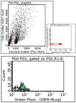
Evaluated by Flow Cytometry in K562 cells.

Flow Cytometry Analysis: 1 µg of this antibody detected IFNAR1 on the surface of K562 cells.

Zielbeschreibung

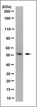
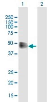
63.53/60.93 kDa (pro-/mature isoform 1), 49.55/46.95 kDa (pro-/mature isoform 2), 56.72/54.12 kDa (pro-/mature isoform 3), 56.05 kDa (isoform 4) calculated. Apparent molecular weight appears greater than 100 kDa due to glycosylation.

Physikalische Form

Protein G purified.
Format: Purified
Purified mouse IgG1κ in buffer containing 0.1 M Tris-Glycine (pH 7.4), 150 mM NaCl with 0.05% sodium azide.

Lagerung und Haltbarkeit

Stable for 1 year at 2-8°C from date of receipt.

Sonstige Hinweise

Concentration: Please refer to lot specific datasheet.

Haftungsausschluss

Unless otherwise stated in our catalog or other company documentation accompanying the product(s), our products are intended for research use only and are not to be used for any other purpose, which includes but is not limited to, unauthorized commercial uses, in vitro diagnostic uses, ex vivo or in vivo therapeutic uses or any type of consumption or application to humans or animals.

Not finding the right product?  

Try our Produkt-Auswahlhilfe.

Lagerklassenschlüssel

12 - Non Combustible Liquids

WGK

WGK 1

Flammpunkt (°F)

Not applicable

Flammpunkt (°C)

Not applicable


Analysenzertifikate (COA)

Suchen Sie nach Analysenzertifikate (COA), indem Sie die Lot-/Chargennummer des Produkts eingeben. Lot- und Chargennummern sind auf dem Produktetikett hinter den Wörtern ‘Lot’ oder ‘Batch’ (Lot oder Charge) zu finden.

Besitzen Sie dieses Produkt bereits?

In der Dokumentenbibliothek finden Sie die Dokumentation zu den Produkten, die Sie kürzlich erworben haben.

Die Dokumentenbibliothek aufrufen

Unser Team von Wissenschaftlern verfügt über Erfahrung in allen Forschungsbereichen einschließlich Life Science, Materialwissenschaften, chemischer Synthese, Chromatographie, Analytik und vielen mehr..

Setzen Sie sich mit dem technischen Dienst in Verbindung.