Pular para o conteúdo
Merck
Todas as fotos(1)

Documentos Principais

MABT840

Sigma-Aldrich

Anti-Myosin-2 (MYH2) Antibody, clone SC-71

clone SC-71, from mouse

Sinônimo(s):

Myosin-2, MyHC-2a, MyHC-IIa, Myosin heavy chain 2, Myosin heavy chain 2a, Myosin heavy chain IIa, Myosin heavy chain, skeletal muscle, adult 2

Faça loginpara ver os preços organizacionais e de contrato


About This Item

Código UNSPSC:
12352203
eCl@ss:
32160702

fonte biológica

mouse

Nível de qualidade

forma do anticorpo

purified immunoglobulin

tipo de produto de anticorpo

primary antibodies

clone

SC-71, monoclonal

reatividade de espécies

human, opossum, rat, mouse

não deve reagir com

guinea pig

reatividade da espécie (prevista por homologia)

bovine (based on 100% sequence homology)

técnica(s)

immunofluorescence: suitable
immunohistochemistry: suitable
western blot: suitable

Isotipo

IgG1κ

nº de adesão NCBI

nº de adesão UniProt

Condições de expedição

ambient

modificação pós-traducional do alvo

unmodified

Informações sobre genes

human ... MYH2(4620)

Descrição geral

Myosin-2 (UniProt Q9BE41; also known as MyHC-2a, MyHC-IIa, Myosin heavy chain 2, Myosin heavy chain 2a, Myosin heavy chain IIa, Myosin heavy chain, skeletal muscle, adult 2) is encoded by the MYHSA gene (Gene ID 788772) in bovine species. Myosin heavy chain (MyHC) is a major structural component of the striated muscle contractile apparatus and is essential for body movement and cardiac contractility. MyHC are encoded by a highly conserved multigene family, of which eight isoforms have been identified in mammals, each encoded by a separate gene that displays distinct temporal-spatial regulation. MyHC-2a is expressed in the fast-type 2A muscle fibers. Recessive MyHC-2a myopathy caused by missense mutations results in mild muscle weakness. Autosomal dominant mutation (E706K) is reported to cause defective muscle function and compromises the structural integrity of all muscle cells. Ref.: Tajsharghi, H et al. (2014). Eur. J. Hum. Genet. 22: 801-808.

Especificidade

Clone SC-71 immunostained type 2A, but not type 1, 2B, or 2X, fibers in rat tibialis anterior muscle by targeting an epitope within the light meromyosin region (Schiaffino, S., et al. (1989). J. Muscle Res. Cell Motil. 10(3):197-205).

Imunogênio

Bovine skeletal muscle myosin (Schiaffino, S., et al. (1989). J. Muscle Res. Cell Motil. 10(3):197-205).

Aplicação

Research Category
Cell Structure
This mouse monoclonal Anti-Myosin-2 (MYH2) Antibody, clone SC-71, Cat. No. MABT840, is validated for use in Immunofluorescence, Immunohistochemistry, and Western Blotting for the detection of Myosin-2.
Western Blotting Analysis: 0.5 µg/mL from a representative lot detected Myosin-2 (MYH2) in 10 µg of human gastrocnemious muscle and showed minimal reactivity with soleus embryonic muscle tissue lysates (Courtesy of Alberto Rossi, Ph.D., University of Colorado, U.S.A.).

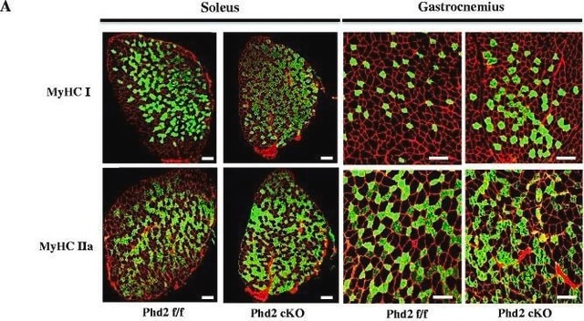
Immunofluorescence Analysis: A representative lot immunostained type 2A fibers in mouse hindlimb muscle cryosections encompassing soleus and surrounding tissues (Kurapati, R., et al. (2012). Hum. Mol. Genet. 21(8):1706-1724).

Immunofluorescence Analysis: A representative lot immunostained type 2A fibers in rat soleus muscle cryosections following Bupivacaine-induced muscle regeneration. In tetrodotoxin/TTX-paralyzed-regenerated muscles type 2A MHC was not expressed (Midrio, M., et al. (2002). Basic Appl. Myol. 12(2): 77-80).

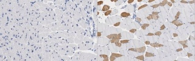
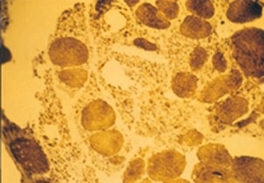
Immunohistochemistry Analysis: A representative lot detected type IIA myosin heavy chain (MyHC) in human masseter (jaw) muscle cryosections (Horton, M.J., et al. (2001). Arch. Oral Biol. 46(11):1039-1050).

Immunohistochemistry Analysis: Representative lots immunostained type 2A, but not type 1, 2B, or 2X, fibers in soleus (rat) and tibialis (mouse, rat, and Mgray short-tailed opossum/Monodelphis domestica) anterior muscle cryosections. Clone SC-71 failed to stain guinea pig tibialis sections (Sciote, J.J., and Rowlerson, A. (1998). Anat. Rec. 251(4):548-562; Gorza, L. (1990). J. Histochem Cytochem. 38(2):257-265; Schiaffino, S., et al. (1989). J. Muscle Res. Cell Motil. 10(3):197-205).

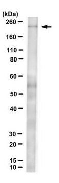
Western Blotting Analysis: A representative lot detected type IIA myosin heavy chain (MyHC) in human masseter (jaw) single muscle fibres extract (Horton, M.J., et al. (2001). Arch. Oral Biol. 46(11):1039-1050).

Western Blotting Analysis: A representative lot detected myosin heavy chain (MHC) in myosin preparations from rat diaphragm, as well as the light meromyosin and rod, but not heavy meromyosin or S-1, fragments of MHC (Schiaffino, S., et al. (1989). J. Muscle Res. Cell Motil. 10(3):197-205).

Qualidade

Identity Confirmation by Isotyping Test.

Isotyping Analysis: The identity of this monoclonal antibody is confirmed by isotyping test to be mouse IgG1 .

Descrição-alvo

~225 kDa observed. 223.3 kDa (bovine), 223.0 (human), 223.2 kDa (mouse) calculated. Uncharacterized bands may be observed in some lysate(s).

forma física

Format: Purified
Protein G purified.
Purified mouse IgG1 in buffer containing 0.1 M Tris-Glycine (pH 7.4), 150 mM NaCl with 0.05% sodium azide.

Armazenamento e estabilidade

Stable for 1 year at 2-8°C from date of receipt.

Outras notas

Concentration: Please refer to lot specific datasheet.

Exoneração de responsabilidade

Unless otherwise stated in our catalog or other company documentation accompanying the product(s), our products are intended for research use only and are not to be used for any other purpose, which includes but is not limited to, unauthorized commercial uses, in vitro diagnostic uses, ex vivo or in vivo therapeutic uses or any type of consumption or application to humans or animals.

Não está encontrando o produto certo?  

Experimente o nosso Ferramenta de seleção de produtos.

Código de classe de armazenamento

12 - Non Combustible Liquids

Classe de risco de água (WGK)

WGK 1


Certificados de análise (COA)

Busque Certificados de análise (COA) digitando o Número do Lote do produto. Os números de lote e remessa podem ser encontrados no rótulo de um produto após a palavra “Lot” ou “Batch”.

Já possui este produto?

Encontre a documentação dos produtos que você adquiriu recentemente na biblioteca de documentos.

Visite a Biblioteca de Documentos

Youngmok C Jang et al.
FASEB journal : official publication of the Federation of American Societies for Experimental Biology, 24(5), 1376-1390 (2009-12-31)
Oxidative stress has been implicated in the etiology of age-related muscle loss (sarcopenia). However, the underlying mechanisms by which oxidative stress contributes to sarcopenia have not been thoroughly investigated. To directly examine the role of chronic oxidative stress in vivo
Brandon M Roberts et al.
FASEB journal : official publication of the Federation of American Societies for Experimental Biology, 27(7), 2600-2610 (2013-03-22)
Cancer cachexia is characterized by a continuous loss of locomotor skeletal muscle mass, which causes profound muscle weakness. If this atrophy and weakness also occurs in diaphragm muscle, it could lead to respiratory failure, which is a major cause of
Diego Salagre et al.
Antioxidants (Basel, Switzerland), 12(8) (2023-08-26)
Obesity-induced skeletal muscle (SKM) inflexibility is closely linked to mitochondrial dysfunction. The present study aimed to evaluate the effects of melatonin on the red vastus lateralis (RVL) muscle in obese rat models at the molecular and morphological levels. Five-week-old male
Tomoya Uchimura et al.
Cell reports. Medicine, 2(6), 100298-100298 (2021-07-02)
Duchenne muscular dystrophy (DMD) is a muscle degenerating disease caused by dystrophin deficiency, for which therapeutic options are limited. To facilitate drug development, it is desirable to develop in vitro disease models that enable the evaluation of DMD declines in contractile

Nossa equipe de cientistas tem experiência em todas as áreas de pesquisa, incluindo Life Sciences, ciência de materiais, síntese química, cromatografia, química analítica e muitas outras.

Entre em contato com a assistência técnica