
Wichtige Dokumente
11296736001
Roche
5-Bromo-2′-deoxy-uridine Labeling and Detection Kit I
sufficient for ≤100 tests, kit of 1 (5 components), suitable for immunofluorescence
Synonym(e):
5-BrdU, 5-Bromo-2-deoxyuridine
About This Item
Empfohlene Produkte


Verwendung
sufficient for ≤100 tests
Qualitätsniveau
Verpackung
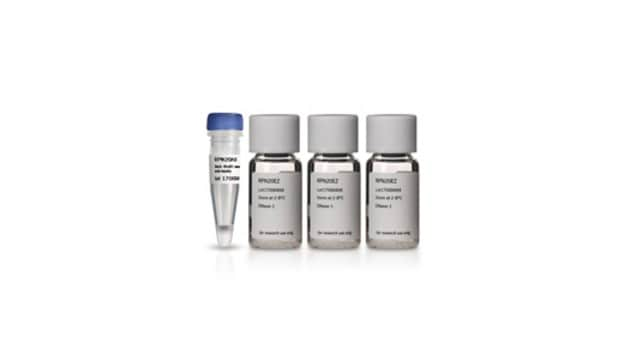
kit of 1 (5 components)
Hersteller/Markenname
Roche
Methode(n)
immunofluorescence: suitable
Lagertemp.
−20°C
Allgemeine Beschreibung
Normally, binding of the antibody is only achieved by denaturation of the DNA. This is usually obtained by exposing the cells to acid, base, or heat. These procedures result in destruction of cell integrity, including cell morphology and surface and cytoplasmatic markers.
The BrdU Labeling and Detection Kit I avoids these problems. The antibody preparation contains specific nucleases which allows access to BrdU after fixation in acidic ethanol. Therefore also simultaneous detection of other markers (double staining) is possible.
Spezifität
Anwendung
- Safe: No radioisotopes are used
- Easy to perform: Follows a standard immunofluorescence protocol
- Sensitive: Denaturation of DNA with nucleases allows for highly sensitive detection of BrdU
- Flexible: Allows double-labeling protocols
BrdU Labeling and Detection Kit has been used for the detection of 5-bromo-2′-deoxy-uridine (BrdU) incorporated into cellular DNA.
Verpackung
Angaben zur Herstellung
Working solution: BrdU labeling medium
Dilute BrdU labeling reagent 1:1000 with sterile cell culture medium (final concentration 10μM).
Note: For in vivo labeling undiluted BrdU labeling reagent (1 to 2ml/100 g body weight) is needed.
Prepare shortly before use.
Anti-BrdU working solution
Dilute anti-BrdU solution 1:10 with Incubation buffer.
Prepare shortly before use.
Anti-mouse-Ig-fluorescein stock solution
Dissolve anti-mouse-Ig-fluorescein solution in 1ml double-dist. water.
Anti-mouse-Ig-fluorescein working solution
Dilute anti-mouse Ig-fluorescein stock solution 1:10 with PBS. If an extended storage is desired, add BSA (bovine serum albumin), 10 mg/ml.
Prepare shortly before use.
Washing buffer
Dilute Washing buffer concentrate (10x) (bottle 2) 1:10 with double-dist. water.
Storage conditions (working solution): BrdU labeling medium
Store undiluted (1000x) medium in aliquots at -15 to -25°C.
Anti-BrdU working solution
Store undiluted antibody at -15 to -25°C.
Anti-mouse-Ig-fluorescein stock solution
Stable at 2 to 8°C
Washing buffer
Stable at 2 to 8°C
Sample material: Cell culture: adherent cells, suspension cells, organ, or explant cultures. Tissue sections (after in vivo labeling with BrdU).
Sonstige Hinweise
Nur Kit-Komponenten
- BrdU Labeling Reagent, sterile 1,000x concentrated
- Washing Buffer concentrate 10x concentrated
- Incubation Buffer
- Anti-BrdU antibody, contains nucleases for DNA denaturation
- Anti-mouse-Ig-fluorescein antibody


Signalwort
Danger
H-Sätze
Gefahreneinstufungen
Aquatic Chronic 3 - Eye Irrit. 2 - Muta. 1B - Skin Irrit. 2 - Skin Sens. 1
Lagerklassenschlüssel
12 - Non Combustible Liquids
WGK
WGK 2
Flammpunkt (°F)
does not flash
Flammpunkt (°C)
does not flash
Hier finden Sie alle aktuellen Versionen:
Besitzen Sie dieses Produkt bereits?
In der Dokumentenbibliothek finden Sie die Dokumentation zu den Produkten, die Sie kürzlich erworben haben.
Kunden haben sich ebenfalls angesehen


Artikel
Zellbasierte Assays für Zellproliferation (BrdU, MTT, WST1), Zellviabilität und Zytotoxizitätsversuche für Anwendungen in der Krebs- und Stammzellforschung und in den Neurowissenschaften.
Cell based assays for cell proliferation (BrdU, MTT, WST1), cell viability and cytotoxicity experiments for applications in cancer, neuroscience and stem cell research.
Unser Team von Wissenschaftlern verfügt über Erfahrung in allen Forschungsbereichen einschließlich Life Science, Materialwissenschaften, chemischer Synthese, Chromatographie, Analytik und vielen mehr..
Setzen Sie sich mit dem technischen Dienst in Verbindung.