跳轉至內容
Merck
全部照片(2)

重要文件

RAB0273

Sigma-Aldrich

人 IL-1 β ELISA 试剂盒

for serum, plasma, cell culture supernatant and urine

同義詞:

Il-1 beta, Interleukin-1 beta

登入查看組織和合約定價


About This Item

分類程式碼代碼:
41116158
NACRES:
NA.84
暫時無法取得訂價和供貨情況

物種活性

human

包裝

kit of 96 wells (12 strips x 8 wells)

技術

ELISA: suitable
capture ELISA: suitable

輸入

sample type urine
sample type serum
sample type plasma
sample type cell culture supernatant(s)

assay range

inter-assay cv: <12%
intra-assay cv: <10%
sensitivity: 0.3 pg/mL
standard curve range: 0.48-100 pg/mL

檢測方法

colorimetric

運輸包裝

wet ice

儲存溫度

−20°C

基因資訊

human ... IL1B(3553)

一般說明

人IL-1 β ELISA(酶联免疫吸附测定)试剂盒采用体外酶联免疫吸附测定法,定量测定人血清、血浆、细胞培养物上清液和尿液中的IL-1 β含量。

免疫原

重组人IL-1β

應用

仅供研究使用。不可用于诊断操作。
人 IL-1 β ELISA试剂盒已被用于测定细胞培养上清液中白细胞介素1β (IL-1 β)的浓度。[1]

生化/生理作用

白细胞介素1β (IL-1 β)是一种促炎细胞因子,其可诱导多种急性和慢性炎症。因此,IL1B被认为是与纤维化和组织重塑相关的疾病的潜在治疗靶标。[2] IL-1β 蛋白会增加内源性92kDa明胶酶(基质金属蛋白酶9(MMP-9))和MMP组织抑制剂(TIMP-1)的产生。[3] 滑液中IL-1β 浓度的升高会促进滑膜炎的发病,以及颞下颌关节软骨组织和骨的退行性变化。[4] IL-1β 可将中性粒细胞和巨噬细胞带到感染部位。[5] IL-1β 基因突变与Henoch-Schönlein紫癜(HSP)的肾脏表现和肾脏后遗症有关。[6]

其他說明

本产品提供样本分析证书。
请在批号对应的文本框中输入单词 sample

套裝中的組件也可單獨購買

產品號碼
描述
SDS

  • RABELADAELISA 1X Assay/Sample Diluent Buffer A (Item D1)SDS

  • RABELADBELISA 5X Assay/Sample Diluent Buffer B (Item E1)SDS

  • RABSTOP3ELISA Stop Solution (Item I)SDS

  • RABTMB3ELISA Colorimetric TMB Reagent (HRP Substrate, Item H)SDS

  • RABWASH420X Wash Buffer (Item B)SDS

象形圖

Corrosion

訊號詞

Warning

危險聲明

防範說明

危險分類

Met. Corr. 1

儲存類別代碼

8A - Combustible corrosive hazardous materials


從最近期的版本中選擇一個:

分析證明 (COA)

Lot/Batch Number

未看到正確版本?

如果您需要一個特定的版本,您可以透過批號來尋找特定憑證。

已經擁有該產品?

您可以在文件庫中找到最近購買的產品相關文件。

存取文件庫

Jie Liu et al.
The Journal of international medical research, 49(3), 300060521996564-300060521996564 (2021-03-27)
Porphyromonas gingivalis (Pg) plays a critical role in the occurrence and development of atherosclerosis. Lipopolysaccharide from Pg (Pg-LPS) could lead to pyroptosis of vascular smooth muscle cells (VSMCs) and induce instability of atherosclerotic plaque. Therefore, pyroptosis of VSMCs could promote
Proinflammatory cytokines detectable in synovial fluids from patients with temporomandibular disorders.
Takahashi T
Oral Surgery, Oral Medicine, Oral Pathology, Oral Radiology, and Endodontics, 85(2), 135-141 (1998)
Zhao Gao et al.
BMC ophthalmology, 20(1), 134-134 (2020-04-08)
Inflammation of RPE cells led to different kinds of eye diseases and affected the normal function of the retina. Furthermore, higher levels of ROCK1 and ROCK2 induced injury of endothelial cells and many inflammatory diseases of the eyes. Ripasudil, which
Zetao Ma et al.
Journal of orthopaedic surgery and research, 15(1), 284-284 (2020-07-30)
Inflammation and apoptosis of chondrocytes are the pathological bases of osteoarthritis. Autophagy could alleviate the symptoms of inflammation and apoptosis. Previous study has shown that BCL2/adenovirus E1B 19 kDa protein-interacting protein 3 (BNIP3) can induce the occurrence and development of autophagy.
Interleukin 1 beta (IL1?) rs16944 genetic variant as a genetic marker of severe renal manifestations and renal sequelae in Henoch-Schonlein purpura.
Lopez-Mejias R
Clinical and Experimental Rheumatology, 34(3 Suppl 97), S84-S88 (2016)

Questions

  1. Human IL-1 β ELISA kit, RAB0273 for this product how to prepare Cell Culture lysate.

    1 answer
    1. This ELISA kit has not been validated for lysates, but it is expected to work. Follow the instructions below for preparing samples.

      Cell Lysates:
      Rinse cells with PBS, ensuring all PBS is removed before detaching cells. Detach 2 x 10^7 cells by trypsinization or scraping and transfer to a microfuge tube. Centrifuge to pellet the cells and remove any remaining buffer. Add approximately 500 µl of prepared lysis buffer and pipette up and down to resuspend the pellet. Incubate the lysates with shaking at 4°C for 30 minutes. Spin down the tubes in a microfuge at top speed (10,000g) for 10 minutes at 4°C, and transfer the supernatants to a clean tube.

      For lysis buffer, use 43-040 Cell Lysis buffer or 20-188 RIPA lysis buffer.
      Guidelines for lysis buffer composition:
      Avoid using more than 0.1% SDS or other strongly denaturing detergents. Non-ionic detergents such as Triton X-100 or NP-40 are recommended, although zwitterionic detergents like CHAPS, or mild ionic detergents such as sodium deoxycholate, will work.
      Use no more than 2% v/v total detergent.
      Avoid sodium azide.
      Avoid using more than 10 mM reducing agents, such as dithiothreitol or mercaptoethanols.
      Adding a protease inhibitor cocktail to the lysis buffer prior to homogenization is strongly recommended. Inhibitor cocktails like P2714 and P8465 can be purchased and used according to their specifications.

      Lysate Application:
      Use cell or tissue lysates immediately or aliquot and store at –80°C. Avoid repeated freeze-thaw cycles. Keep thawed lysates on ice prior to use. For the first experiment, perform serial dilution testing, starting with a 5-fold dilution, to determine the optimal protein load for the assay. Optimal dilution depends on the abundance of target proteins and should be determined empirically. Determine the total protein concentration using the Pierce BCA Protein Assay Kit, Cat#: 23227. Dilute lysate to a final total protein concentration of 50-500 µg/ml (5-fold or more is preferred) when performing antibody array or ELISA testing.
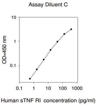
      Dilute the cell lysate for this kit with Assay Diluent B. For other ELISA kits validated with cell lysate samples, a minimum 5-fold dilution is recommended to avoid sample matrix effects, but the optimal sample dilution must be determined empirically by the researcher.

      Helpful?

Reviews

No rating value

Active Filters

我們的科學家團隊在所有研究領域都有豐富的經驗,包括生命科學、材料科學、化學合成、色譜、分析等.

聯絡技術服務