
推薦產品


生物源
rabbit
品質等級
共軛
unconjugated
抗體表格
affinity isolated antibody
抗體產品種類
primary antibodies
無性繁殖
polyclonal
產品線
Prestige Antibodies® Powered by Atlas Antibodies
形狀
buffered aqueous glycerol solution
物種活性
human
技術
immunofluorescence: 0.25-2 μg/mL
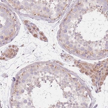
immunohistochemistry: 1:200-1:500
免疫原序列
LKRLQEKVEVLEAKKEELETENQVLNRQNVPFEDYTRLQKRLKDIQRRHNEFRSLILVPNMPPTASINPVSFQSSAMVPSMELPFPPHMQEEQHQRELSL
運輸包裝
wet ice
儲存溫度
−20°C
目標翻譯後修改
unmodified
基因資訊
human ... CCDC41(51134)
一般說明
The gene CEP83 (centrosomal protein 83) encodes a protein with coiled-coil domains.
免疫原
centrosomal protein 83kDa
應用
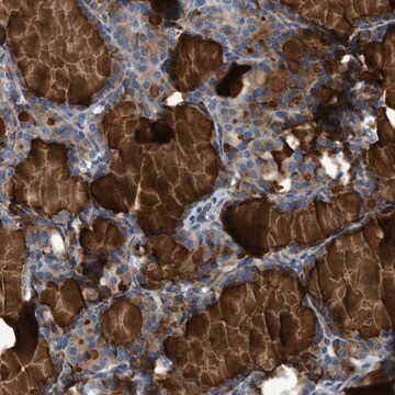
All Prestige Antibodies®Powered by Atlas Antibodies is developed and validated by the Human Protein Atlas (HPA) project . Each antibody is tested by immunohistochemistry against hundreds of normal and disease tissues. These images can be viewed on the Human Protein Atlas (HPA) site by clicking on the Image Gallery link. We also provide Prestige Antibodies® protocols and other useful information.
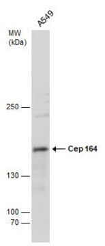
Anti-CEP83 antibody produced in rabbit has been used in western blotting and immunofluorescence.
Anti-CEP83 antibody produced in rabbit has been used in western blotting and immunofluorescence.
生化/生理作用
CEP83 (centrosomal protein 83) plays an important role in ciliogenesis. It is part of DAPs (distal appendages) in centrioles. In ciliogenesis, DAPs are needed for docking and attachment of the mother centriole to the cellular surface. CEP83 is also required for bringing other components (for instance CEP164) to the mother centriole. Mutation in this gene is associated with early-onset infantile nephronophthisis and intellectual disability.
特點和優勢
Prestige Antibodies® are highly characterized and extensively validated antibodies with the added benefit of all available characterization data for each target being accessible via the Human Protein Atlas portal linked just below the product name at the top of this page. The uniqueness and low cross-reactivity of the Prestige Antibodies® to other proteins are due to a thorough selection of antigen regions, affinity purification, and stringent selection. Prestige antigen controls are available for every corresponding Prestige Antibody and can be found in the linkage section.
Every Prestige Antibody is tested in the following ways:
Every Prestige Antibody is tested in the following ways:
- IHC tissue array of 44 normal human tissues and 20 of the most common cancer type tissues.
- Protein array of 364 human recombinant protein fragments.
聯結
Corresponding Antigen APREST81162
外觀
Solution in phosphate buffered saline, pH 7.2, containing 40% glycerol and 0.02% sodium azide.
法律資訊
Prestige Antibodies is a registered trademark of Merck KGaA, Darmstadt, Germany
免責聲明
Unless otherwise stated in our catalog or other company documentation accompanying the product(s), our products are intended for research use only and are not to be used for any other purpose, which includes but is not limited to, unauthorized commercial uses, in vitro diagnostic uses, ex vivo or in vivo therapeutic uses or any type of consumption or application to humans or animals.
未找到適合的產品?
試用我們的產品選擇工具.
儲存類別代碼
10 - Combustible liquids
水污染物質分類(WGK)
WGK 1
閃點(°F)
Not applicable
閃點(°C)
Not applicable
Lauren T Evans et al.
The EMBO journal, 40(4), e105106-e105106 (2020-12-23)
Centriole copy number is tightly maintained by the once-per-cycle duplication of these organelles. Centrioles constitute the core of centrosomes, which organize the microtubule cytoskeleton and form the poles of the mitotic spindle. Centrosome amplification is frequently observed in tumors, where
SDCCAG8 Interacts with RAB Effector Proteins RABEP2 and ERC1 and Is Required for Hedgehog Signaling.
Rannar Airik et al.
PloS one, 11(5), e0156081-e0156081 (2016-05-26)
Recessive mutations in the SDCCAG8 gene cause a nephronophthisis-related ciliopathy with Bardet-Biedl syndrome-like features in humans. Our previous characterization of the orthologous Sdccag8gt/gt mouse model recapitulated the retinal-renal disease phenotypes and identified impaired DNA damage response signaling as an underlying
Mathew Bowler et al.
Nature communications, 10(1), 993-993 (2019-03-03)
Centrioles are vital cellular structures that form centrosomes and cilia. The formation and function of cilia depends on a set of centriole's distal appendages. In this study, we use correlative super resolution and electron microscopy to precisely determine where distal
Bahtiyar Kurtulmus et al.
Journal of cell science, 131(18) (2018-08-23)
Cilia perform essential signalling functions during development and tissue homeostasis. A key event in ciliogenesis occurs when the distal appendages of the mother centriole form a platform that docks ciliary vesicles and removes CP110-Cep97 inhibitory complexes. Here, we analysed the
Noémie Gaudin et al.
eLife, 11 (2022-03-24)
Centrioles are formed by microtubule triplets in a ninefold symmetric arrangement. In flagellated protists and animal multiciliated cells, accessory structures tethered to specific triplets render the centrioles rotationally asymmetric, a property that is key to cytoskeletal and cellular organization in
我們的科學家團隊在所有研究領域都有豐富的經驗,包括生命科學、材料科學、化學合成、色譜、分析等.
聯絡技術服務