跳轉至內容
Merck
全部照片(3)

文件

C1801

Sigma-Aldrich

抗细胞角蛋白单抗 小鼠抗

clone PCK-26, ascites fluid

同義詞:

登入查看組織和合約定價


About This Item

分類程式碼代碼:
12352203
NACRES:
NA.41

生物源

mouse

品質等級

共軛

unconjugated

抗體表格

ascites fluid

抗體產品種類

primary antibodies

無性繁殖

PCK-26, monoclonal

包含

15 mM sodium azide

物種活性

human, chicken, snake, hamster, pig, goat, feline, bovine, carp, rat, rabbit, canine, lizard, sheep, mouse, guinea pig

技術

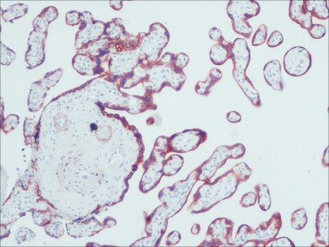
immunohistochemistry (formalin-fixed, paraffin-embedded sections): 1:300
immunohistochemistry (frozen sections): suitable
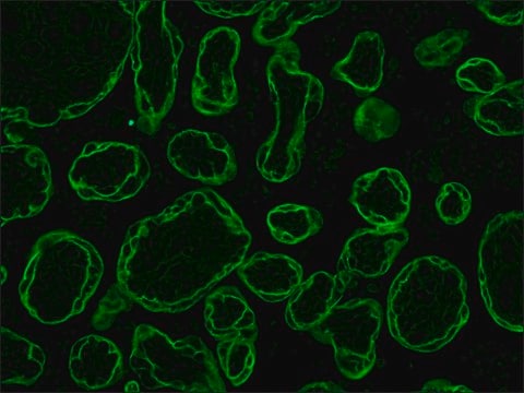
indirect immunofluorescence: 1:300 using protease-idgested, formalin-fixed, paraffin-embedded sections of human or animal tissues
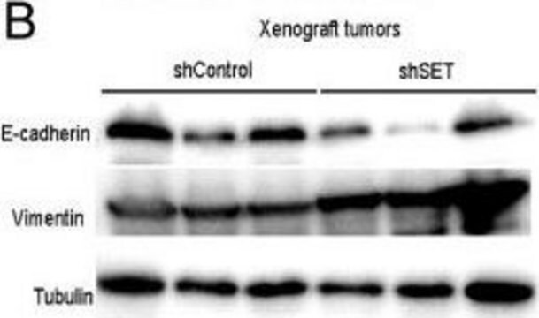
western blot: suitable

同型

IgG1

運輸包裝

dry ice

儲存溫度

−20°C

目標翻譯後修改

unmodified

尋找類似的產品? 前往 產品比較指南

一般說明

细胞角蛋白属于中间丝(IF)蛋白家族的成员。
细胞角蛋白,包括至少29种不同的蛋白质,是上皮细胞和单核细胞的特征蛋白。 细胞角蛋白1、5、6和8是II型中性至碱性亚家族的成员。细胞角蛋白肽1(68 kDa)与细胞角蛋白10一起在基底上层细胞层或表皮分化区室中表达。它的表达随着表皮的成熟而增加,并且在角质层的终末分化角质形成细胞中被翻译后修饰。 细胞角蛋白肽5(58 kDa)是分层上皮细胞中的主要II型角蛋白,而细胞角蛋白8型(52 kDa)是简单上皮细胞中的主要II型角蛋白。 细胞角蛋白6(56 kDa)是在具有自然或病理性高周转率的组织中表达的“过度增殖”细胞角蛋白。

特異性

该抗体可识别位于II型细胞角蛋白1、5、6和8上的表位。PCK-26是一种广谱抗体,可与多种正常、反应性和赘生性上皮组织发生特异性反应。该抗体可与简单、成角和非成角的鳞状上皮和假复层上皮反应。

免疫原

来源于人表皮的细胞角蛋白。

應用

抗细胞角蛋白的单克隆抗体是一种上皮细胞分化的特异性标志物,已被广泛用作肿瘤鉴定和分类的工具。 单克隆抗细胞角蛋白是一种广泛反应的基团型抗体,可识别大多数人上皮组织中存在的表位。 它有助于正常细胞、化生细胞和赘生性细胞的分型,并有助于区分癌瘤和非上皮性肿瘤,例如肉瘤、淋巴瘤和神经肿瘤。 它也可用于检测淋巴结和其他组织中的微转移以及确定低分化肿瘤的起源。
单克隆抗细胞角蛋白可以用于通过各种免疫化学测定(例如免疫印迹、斑点印迹和免疫组织化学(免疫荧光和免疫酶染色))定位细胞角蛋白。
通过对人体或动物组织进行蛋白酶消化、福尔马林固定、石蜡包埋,并对切片进行间接免疫荧光染色,确定了其最小抗体滴度为1:300。

生化/生理作用

全细胞角蛋白以及CXCR4的表达可能是非小细胞肺癌(NSCLC)预后的生物标志物。

免責聲明

除非我们的产品目录或产品附带的其他公司文档另有说明,否则我们的产品仅供研究使用,不得用于任何其他目的,包括但不限于未经授权的商业用途、体外诊断用途、离体或体内治疗用途或任何类型的消费或应用于人类或动物。

Not finding the right product?  

Try our 產品選擇工具.

儲存類別代碼

10 - Combustible liquids

水污染物質分類(WGK)

WGK 3

閃點(°F)

Not applicable

閃點(°C)

Not applicable


分析證明 (COA)

輸入產品批次/批號來搜索 分析證明 (COA)。在產品’s標籤上找到批次和批號,寫有 ‘Lot’或‘Batch’.。

已經擁有該產品?

您可以在文件庫中找到最近購買的產品相關文件。

存取文件庫

Christopher W Helsen et al.
Nature communications, 9(1), 3049-3049 (2018-08-05)
Engineering T cells with chimeric antigen receptors (CARs) is an effective method for directing T cells to attack tumors, but may cause adverse side effects such as the potentially lethal cytokine release syndrome. Here the authors show that the T
Clinical utility of cytokeratins as tumor markers
Barak V, et al.
Clinical Biochemistry, 37(7), 529-540 (2004)
Anup Sood et al.
Viruses, 12(8) (2020-07-29)
Over the last 15 years, advances in immunofluorescence-imaging based cycling methods, antibody conjugation methods, and automated image processing have facilitated the development of a high-resolution, multiplexed tissue immunofluorescence (MxIF) method with single cell-level quantitation termed Cell DIVETM. Originally developed for
Marcus Ruscetti et al.
Cancer research, 75(13), 2749-2759 (2015-05-08)
The epithelial-mesenchymal transition (EMT) has been postulated as a mechanism by which cancer cells acquire the invasive and stem-like traits necessary for distant metastasis. However, direct in vivo evidence for the role of EMT in the formation of cancer stem-like
Charles A Herring et al.
Cell systems, 6(1), 37-51 (2017-11-21)
Modern single-cell technologies allow multiplexed sampling of cellular states within a tissue. However, computational tools that can infer developmental cell-state transitions reproducibly from such single-cell data are lacking. Here, we introduce p-Creode, an unsupervised algorithm that produces multi-branching graphs from

我們的科學家團隊在所有研究領域都有豐富的經驗,包括生命科學、材料科學、化學合成、色譜、分析等.

聯絡技術服務