MABF125
Anti-TAP1 Antibody, clone mAb 148.3
clone mAb 148.3, from mouse
别名:
Antigen peptide transporter 1, APT1, ATP-binding cassette sub-family B member 2, Peptide supply factor 1, Peptide transporter PSF1, PSF-1, Peptide transporter TAP1, Peptide transporter involved in antigen processing 1, Really interesting new gene 4 prote
登录查看公司和协议定价
所有图片(1)
About This Item
分類程式碼代碼:
12352203
eCl@ss:
32160702
NACRES:
NA.41
推荐产品
生物源
mouse
品質等級
抗體表格
purified immunoglobulin
抗體產品種類
primary antibodies
無性繁殖
mAb 148.3, monoclonal
物種活性
human
技術
activity assay: suitable
immunofluorescence: suitable
immunoprecipitation (IP): suitable
western blot: suitable
同型
IgG1κ
NCBI登錄號
UniProt登錄號
運輸包裝
wet ice
目標翻譯後修改
unmodified
基因資訊
human ... TAP1(6890)
一般說明
Peptide transporter TAP1 is also called Antigen peptide transporter 1 (APT1), ATP-binding cassette sub-family B member 2 (ABCB2), Peptide supply factor 1 (PSF1), Peptide transporter PSF1 (PSF-1), Peptide transporter involved in antigen processing 1, and Really interesting new gene 4 protein (RING4). TAP1 is involved in the transport of antigens from the cytoplasm to the endoplasmic reticulum for association with MHC class I molecules as well as MHC class I folding. TAP1 is inhibited herpes simplex virus ICP47 protein, human cytomegalovirus US6 glycoprotein, human adenovirus E3-19K glycoprotein, and is down-regulated by human Epstein-Barr virus vIL-10 protein. TAP1 mutations can be a cause of Bare lymphocyte syndrome 1 (BLS1).
免疫原
Epitope: This antibody recognizes the ADAPE amino acid residues within the CYWAMVQAPADAPE sequence of human TAP1 (located at the C-terminus).
Linear peptide corresponding to the CYWAMVQAPADAPE sequence of human TAP1.
應用
Detect Antigen peptide transporter 1 using this mouse monoclonal antibody, Anti-TAP1 Antibody, clone mAb 148.3 validated for use in western blotting, IP, Immunofluorescence & Activity Assay.
Western Blotting Analysis: A representative lot from an independent laboratory detected TAP1 in transiently transfected HeLa cells (Hulpke, S., et al. (2012). FASEB J. 26(12):5071-5080.).
Western Blotting Analysis: A representative lot from an independent laboratory detected TAP1 in microsomes of baculovirus-infected SF9 cells, which express wild type or select mutations of TAP1 (Chen, M., et al. (2004). J Biol Chem. 279(44):46073-46081.).
Western Blotting Analysis: A representative lot from an independent laboratory detected TAP1 in SF9 cells infected with recombinant baculovirus containing TAP1 gene constructs (Meyer, T. H., et al. (1994). FEBS Lett. 351(3):443-447.).
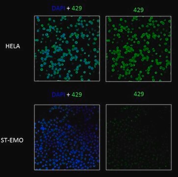
Immunofluorescence Analysis: A representative lot from an independent laboratory detected TAP1 in HeLa cells contransfected with wild type TAP1 and TAP2 (Hulpke, S., et al. (2012). Cell Mol Life Sci. 69(19):3317-3327.).
Immunofluorescence Analysis: A representative lot from an independent laboratory detected TAP1 in SF9 cells infected with rBV-TAP1/rBV-TAP2 (Meyer, T. H., et al. (1994). FEBS Lett. 351(3):443-447.).
Immunoprecipitation Analysis: A representative lot from an independent laboratory immunoprecipitated TAP1 from SF9 cells infected with rBV-TAP1/rBV-TAP2 (Meyer, T. H., et al. (1994). FEBS Lett. 351(3):443-447.).
Activity Assay Analysis: This antibody inhibits TAP-specific peptide transport (Plewnia, G., et al. (2007). J Mol Biol. 369(1):95-107.).
品質
Evaluated by Western Blotting in untreated and Interferon-gamma (IFN-g) treated HeLa cell lysate.
Western Blotting Analysis: 0.5 µg/mL of this antibody detected TAP1 in 10 µg of Interferon-gamma (IFN-g) treated HeLa cell lysate.
Western Blotting Analysis: 0.5 µg/mL of this antibody detected TAP1 in 10 µg of Interferon-gamma (IFN-g) treated HeLa cell lysate.
標靶描述
~65 kDa observed. Uniprot describes a molecular weight of ~87 kDa However, this protein may be observed at ~65 kDa (Koch, J., et al. (2006). FEBS Lett. 580(17):4091-4096.).
外觀
Format: Purified
Purified mouse monoclonal IgG1κ supernatant in buffer containing PBS without preservatives.
其他說明
Concentration: Please refer to the Certificate of Analysis for the lot-specific concentration.
未找到合适的产品?
试试我们的产品选型工具.
儲存類別代碼
12 - Non Combustible Liquids
水污染物質分類(WGK)
WGK 2
閃點(°F)
Not applicable
閃點(°C)
Not applicable
Man Huang et al.
Scientific reports, 6, 33612-33612 (2016-09-17)
HLA class I (HLA-I) transgenic mice have proven to be useful models for studying human MHC-related immune responses over the last two decades. However, differences in the processing and presentation machinery between humans and mice may have profound effects on
Brendan L C Kinney et al.
Cancer immunology, immunotherapy : CII, 73(1), 10-10 (2024-01-17)
The antigen processing machinery (APM) components needed for a tumor cell to present an antigen to a T cell are expressed at low levels in solid tumors, constituting an important mechanism of immune escape. More than most other solid tumors
Ilse Dingjan et al.
European journal of cell biology, 96(7), 705-714 (2017-07-10)
Cross-presentation of foreign antigen in major histocompatibility complex (MHC) class I by dendritic cells (DCs) requires activation of the NADPH-oxidase NOX2 complex. We recently showed that NOX2 is recruited to phagosomes by the SNARE protein VAMP8 where NOX2-produced reactive oxygen
Devin Dersh et al.
Immunity, 54(1), 116-131 (2020-12-04)
Tumors frequently subvert major histocompatibility complex class I (MHC-I) peptide presentation to evade CD8+ T cell immunosurveillance, though how this is accomplished is not always well defined. To identify the global regulatory networks controlling antigen presentation, we employed genome-wide screening in
我们的科学家团队拥有各种研究领域经验,包括生命科学、材料科学、化学合成、色谱、分析及许多其他领域.
联系技术服务部门


