推荐产品
生物源
rabbit
品質等級
抗體表格
purified antibody
抗體產品種類
primary antibodies
無性繁殖
polyclonal
物種活性
mouse, human
技術
immunocytochemistry: suitable
immunoprecipitation (IP): suitable
western blot: suitable
NCBI登錄號
UniProt登錄號
運輸包裝
wet ice
目標翻譯後修改
unmodified
基因資訊
human ... SH3PXD2A(9644)
mouse ... Sh3Pxd2A(14218)
一般說明
Tks5(以前称为Fish)是一种大型支架蛋白,具有一个氨基末端PX结构域和五个SH3结构域。它在正常成纤维细胞中是细胞质的。在Src转化的细胞中,Tks5定位于足小体。Tks5和组成型活性Src在上皮癌细胞中的共表达导致足小体/侵袭性伪足的出现。Tks5表达降低的细胞通过基质胶的侵袭性很差。Tks5在一些侵袭性人类癌细胞系和肿瘤组织中表达并定位于足小体/侵袭性伪足,特别是乳腺癌和黑色素瘤。因此,Tks5似乎是足小体/侵袭性伪足形成和某些癌细胞侵袭所必需的。
特異性
其他同源性:人、犬、马、大鼠和牛(100%序列同源性),鸡(93%序列同源性)
免疫原
表位:166-225 aa
重组蛋白,aa:166-225。同源性:人、犬、马、大鼠、牛(100%,60/60);鸡(93%,56/60)
應用
抗TKS5 (SH3 #1)抗体可检测TKS5 (SH3 #1) 水平 & 已出版&并经过验证可用于WB,IC & IP。
研究子类别
细胞骨架信号转导
细胞骨架信号转导
研究类别
细胞结构
细胞结构
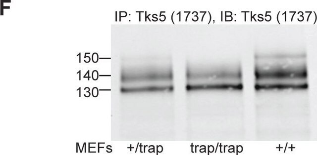
蛋白质印迹分析:抗体可在20 μg NIH/3T3(100%融合)裂解液中检测到TKS5。
品質
通过蛋白质印迹在NIH/3T3(100%融合)裂解液中进行了常规评估。
標靶描述
130-150 kDa 某些细胞裂解液中可能会出现未表征的条带。
外觀
在含0.05%NaN3的0.1M Tris-甘氨酸(pH7.4)150mM NaCl中的纯化抗体。
形式:纯化
蛋白A纯化
儲存和穩定性
自收到之日起,在2-8°C以未稀释的等分试样可保存1年。
分析報告
对照
蛋白质印迹:
NIH/3T3(100%融合)裂解液
免疫细胞化学:
小鼠3T3-Src(Y527F)细胞
蛋白质印迹:
NIH/3T3(100%融合)裂解液
免疫细胞化学:
小鼠3T3-Src(Y527F)细胞
其他說明
浓度:请参考批次特异性浓缩物的分析证书。
免責聲明
除非我们的产品目录或产品附带的其他公司文档另有说明,否则我们的产品仅供研究使用,不得用于任何其他目的,包括但不限于未经授权的商业用途、体外诊断用途、离体或体内治疗用途或任何类型的消费或应用于人类或动物。
未找到合适的产品?
试试我们的产品选型工具.
儲存類別代碼
12 - Non Combustible Liquids
水污染物質分類(WGK)
WGK 1
閃點(°F)
Not applicable
閃點(°C)
Not applicable
Samuel J Ghilardi et al.
Molecular biology of the cell, 32(18), 1707-1723 (2021-07-01)
Interactions between the actin cytoskeleton and the plasma membrane are important in many eukaryotic cellular processes. During these processes, actin structures deform the cell membrane outward by applying forces parallel to the fiber's major axis (as in migration) or they
Karla C Williams et al.
Oncogene, 38(19), 3598-3615 (2019-01-18)
Invadopodia are cell protrusions that mediate cancer cell extravasation but the microenvironmental cues and signaling factors that induce invadopodia formation during extravasation remain unclear. Using intravital imaging and loss of function experiments, we determined invadopodia contain receptors involved in chemotaxis
Nawal Bendris et al.
Journal of cell science, 129(14), 2804-2816 (2016-06-10)
The ability of cancer cells to degrade the extracellular matrix and invade interstitial tissues contributes to their metastatic potential. We recently showed that overexpression of sorting nexin 9 (SNX9) leads to increased cell invasion and metastasis in animal models, which
Deciphering the involvement of the Hippo pathway co-regulators, YAP/TAZ in invadopodia formation and matrix degradation.
Venghateri, et al.
Cell Death & Disease, 14, 290-290 (2023)
Rac3 regulates breast cancer invasion and metastasis by controlling adhesion and matrix degradation.
Sara K Donnelly et al.
The Journal of cell biology, 216(12), 4331-4349 (2017-10-25)
The initial step of metastasis is the local invasion of tumor cells into the surrounding tissue. Invadopodia are actin-based protrusions that mediate the matrix degradation necessary for invasion and metastasis of tumor cells. We demonstrate that Rac3 GTPase is critical
我们的科学家团队拥有各种研究领域经验,包括生命科学、材料科学、化学合成、色谱、分析及许多其他领域.
联系技术服务部门


