콘텐츠로 건너뛰기
Merck
모든 사진(1)

주요 문서

MAB351R

Sigma-Aldrich

Anti-GAD2(GAD67) Antibody

CHEMICON®, mouse monoclonal, GAD-6

동의어(들):

GAD65

로그인조직 및 계약 가격 보기

크기 선택

100 μG
₩804,286

₩804,286


예상 입고일2025년 5월 12일세부사항


벌크 견적 요청

크기 선택

보기 변경
100 μG
₩804,286

About This Item

UNSPSC 코드:
12352203
eCl@ss:
32160702
NACRES:
NA.41

₩804,286


예상 입고일2025년 5월 12일세부사항


벌크 견적 요청

제품명

Anti-Glutamate Decarboxylase Antibody, 65 kDa isoform, clone GAD-6, clone GAD-6, Chemicon®, from mouse

생물학적 소스

mouse

Quality Level

항체 형태

purified immunoglobulin

항체 생산 유형

primary antibodies

클론

GAD-6, monoclonal

종 반응성

human, rat

제조업체/상표

Chemicon®

기술

immunohistochemistry: suitable
western blot: suitable

동형

IgG2a

NCBI 수납 번호

UniProt 수납 번호

배송 상태

dry ice

타겟 번역 후 변형

unmodified

유전자 정보

human ... GAD2(2572)

일반 설명

Gutamic acid decarboxylase (GAD; E.C. 4.1.1.15) is the enzyme responsible for the conversion of glutamic acid to gamma-aminobutyric acid (GABA), the major inhibitory transmitter in higher brain regions, and putative paracrine hormone in pancreatic islets. Two molecular forms of GAD (65kDa and 67kDa, 64% aa identity between forms) are highly conserved and both forms are expressed in the CNS, pancreatic islet cells, testis, oviduct and ovary. The isoforms are regionally distributed cytoplasmically in the brains of rats and mice (Sheikh, S. et al. 1999). GAD65 is an ampiphilic, membrane-anchored protein (585aa), encoded on human chromosome 10, and is responsible for vesicular GABA production. GAD67 is cytoplasmic (594aa.), encoded on chromosome 2, and seems to be responsible for significant cytoplasmic GABA production. GAD expression changes during neural development in rat spinal cord. GAD65 is expressed transiently in commissural axons around E13 but is down regulated the next day while GAD67 expression increases mostly in the somata of those neurons (Phelps, P. et al. 1999). In mature rat pancreas, GAD65 and GAD67 appear to be differentially localized, GAD65 primarily in insulin-containing beta cells and GAD67 in glucagon-containing (A) cells (Li, L. et al. 1995). GAD67 expression seems to be particularly plastic and can change in response to experimental manipulation (for example neuronal stimulation or transection) or disease progression and emergent disorders like schizophrenia (Volk et al., 2000). Colocalization of the two GAD isoforms also shows changes in GAD65/GAD67 distributions correlated with certain disease states such as IDDM and SMS.

특이성

Recognizes the lower molecular weight isoform of the two GAD isoforms identified in brain (Gottlieb, et al., 1986; Chang & Gottlieb, 1988). This monclonal antibody can be used for immunohistochemical localization in brain or pancreas. Anti-GAD has also been used to label purified GAD on Western blots (Chang & Gottlieb, 1988).

면역원

Purified rat brain GAD.

애플리케이션

Anti-Glutamate Decarboxylase Antibody, 65 kDa isoform, clone GAD-6 is an antibody against Glutamate Decarboxylase for use in IH & WB.
Immunohistochemistry: (≤ 1 μg/ml) Optimal working dilutions must be determined by end user.


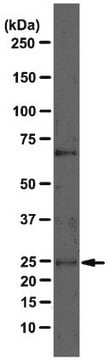
Immunohystochemical Staining Procedures

The following procedure was developed to localize GAD in rat brain sections of cerebellum. Perform all steps at room temperature unless otherwise indicated. Where normal serum is indicated, use normal serum from the same species as the source of the secondary antibody.This procedure represents suggested guidelines for the use of anti-GAD. Fixation regimen, antibody concentrations, and incubation conditions for a given experimental system should be determined empirically.

1. Perfuse rats with 100 mM phosphate buffer, pH 7.4, containing 1% paraformaldehyde, 0.34% L-lysine, and 0.05% sodium m-periodate (1% PLP).

2. Postfix brains in 1% PLP for 1-2 hours. Longer fixation times may reduce labeling intensity.

3. Transfer brains to 100 mM phosphate buffer containing 30% sucrose, and gently agitate on a shaker platform at +4°C for 48-60 hours.

4. Using a sliding microtome, cut 30 mm sections of frozen cerebellum. As the sections are cut, collect them in a vial of cold 100 mM phosphate buffer.

5. Incubate sections in phosphate-buffered saline (PBS) containing 1.5% normal serum and 0.2% TritonX-100 for 30 minutes.

6. On a shaker platform, incubate sections with anti-GAD (diluted in PBS containing 1.5% normal serum and 0.2% Triton X-100 to a final antibody concentration of 1 mg/ml) for 12-36 hours at +4°C.

7. On a shaker platform, rinse sections eight times, 10-15 minutes per rinse, in PBS.

8. Detect with a standard secondary antibody detection system (Hsu et al., 1981; Falini & Taylor, 1983; Harlow & Lane, 1988; Taylor, 1978).

9. Mount sections, dehydrate, and apply coverslips.
Research Category
Neuroscience
Research Sub Category
Neurotransmitters & Receptors

표적 설명

65 kDa

물리적 형태

Ammonium sulfate precipitation and DEAE-cellulose chromatography
Format: Purified
Lyophilized. Dissolve contents of vial in 100 µL of sterile, distilled water. This results in a final antibody concentration of 1 mg/ml in 10 mM potassium phosphate, 70 mM sodium chloride, pH 7.4 containing no preservatives.

저장 및 안정성

Maintain for 1 year at -20°C from date of shipment. Aliquot to avoid repeated freezing and thawing. For maximum recovery of product, centrifuge the original vial after thawing and prior to removing the cap.

분석 메모

Control
Brain tissue

기타 정보

Concentration: Please refer to the Certificate of Analysis for the lot-specific concentration.

법적 정보

CHEMICON is a registered trademark of Merck KGaA, Darmstadt, Germany

면책조항

Unless otherwise stated in our catalog or other company documentation accompanying the product(s), our products are intended for research use only and are not to be used for any other purpose, which includes but is not limited to, unauthorized commercial uses, in vitro diagnostic uses, ex vivo or in vivo therapeutic uses or any type of consumption or application to humans or animals.

적합한 제품을 찾을 수 없으신가요?  

당사의 제품 선택기 도구.을(를) 시도해 보세요.

픽토그램

Exclamation mark

신호어

Warning

유해 및 위험 성명서

Hazard Classifications

Acute Tox. 4 Dermal - Acute Tox. 4 Inhalation - Acute Tox. 4 Oral - Aquatic Chronic 3

Storage Class Code

13 - Non Combustible Solids

WGK

WGK 3

Flash Point (°F)

Not applicable

Flash Point (°C)

Not applicable


시험 성적서(COA)

제품의 로트/배치 번호를 입력하여 시험 성적서(COA)을 검색하십시오. 로트 및 배치 번호는 제품 라벨에 있는 ‘로트’ 또는 ‘배치’라는 용어 뒤에서 찾을 수 있습니다.

이 제품을 이미 가지고 계십니까?

문서 라이브러리에서 최근에 구매한 제품에 대한 문서를 찾아보세요.

문서 라이브러리 방문

Distribution of alpha1, alpha4, gamma2, and delta subunits of GABAA receptors in hippocampal granule cells.
Sun, C; Sieghart, W; Kapur, J
Brain Research null
Lorenzo Gesuita et al.
Cell reports, 40(7), 111209-111209 (2022-08-18)
Microglia play a key role in shaping the formation and refinement of the excitatory network of the brain. However, less is known about whether and how they organize the development of distinct inhibitory networks. We find that microglia are essential
Chenying Guo et al.
The Journal of comparative neurology, 518(10), 1647-1669 (2010-03-18)
Gamma-aminobutyric acid (GABA) is likely expressed in horizontal cells of all species, although conflicting physiological findings have led to considerable controversy regarding its role as a transmitter in the outer retina. This study has evaluated key components of the GABA
A shared vesicular carrier allows synaptic corelease of GABA and glycine.
Wojcik, SM; Katsurabayashi, S; Guillemin, I; Friauf, E; Rosenmund, C; Brose, N; Rhee, JS
Neuron null
Yoav Noam et al.
The Journal of comparative neurology, 520(13), 3013-3034 (2012-03-22)
Filamin A (FLNa) is an actin-binding protein that regulates cell motility, adhesion, and elasticity by cross-linking filamentous actin. Additional roles of FLNa include regulation of protein trafficking and surface expression. Although the functions of FLNa during brain development are well

문서

Human iPSC neural differentiation media and protocols used to generate neural stem cells, neurons and glial cell types.

질문

후기

평점 값 없음

활성 필터

자사의 과학자팀은 생명 과학, 재료 과학, 화학 합성, 크로마토그래피, 분석 및 기타 많은 영역을 포함한 모든 과학 분야에 경험이 있습니다..

고객지원팀으로 연락바랍니다.